amnion - flat, not columnar epithelium, pathologic if columnar
amniotic sac
chorion
trophoblast differentiate into cytotrophoblast and syncytiotrophoblast
cotyledons(?)
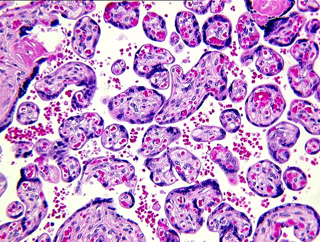
syncytial knots
remnants of allantoic duct(distal) lined by flat or cuboidal cells, omphalomesenteric duct(peripheral) by columnar cells with occasionally goblet cells
vimentin, immunoperoxidase, cytokeratin
oligohydramninos, polyhydramninos
Potter syndrome due to fetal kidney cyst - oligoid fluid
fetal demise - hydrops fetalis, cystic hygroma
nucleated blood cell - due to stress in placenta
preeclampsia, eclampsia - magnesium sulfate lowers BP in preeclampsia
line of Zahnn
PROM (premature rupture of membrane)
breech birth
placenta previa
vasa previa
placental abruption
meconium - abundance may be sign of acidosis
APGAR
--------------------------------------------------------
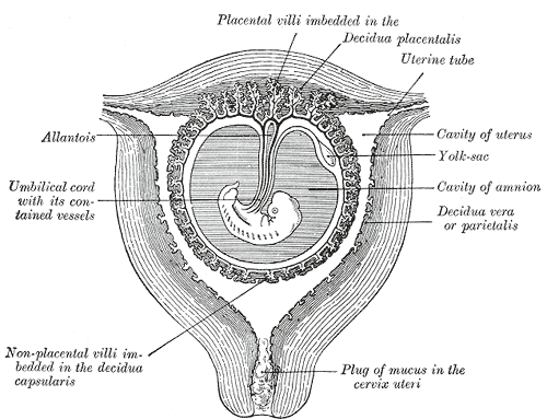
Decidua is the term for the uterine lining (endometrium) during a pregnancy. It is formed under the influence of progesterone and serves to support and interact with the gestation. The decidua represents the maternal portion of the placenta.
Uterine Mucous Membrane

Background
After ovulation, in mammals, the endometrial lining becomes transformed into a secretory lining in preparation to accept the embryo. Without implantation the secretory lining will be absorbed (estrous cycle) or shed (menstrual cycle).
With implantation the lining now termed decidua evolves further during the pregnancy.
The decidua is shed during the parturition process.
[edit] Structure
Different layers of the deciduas have been described:
- a compact outer layer (stratum compactum)
- an intermediate layer (stratum spongiosum)
- a boundary layer adjacent to the myometrium
That part of the deciduas that interacts with the trophoblast is the decidua vera (“true decidua”). The remainder of the decidua is termed the decidua parietalis.
The decidua has a histologically distinct appearance displaying large polygonal decidual cells in the stroma.
Formation of a specialized decidua is called decidualization which is a special property of endometrium seen only in hemochorial placentation.
Decidualization includes the process of differentiation of the spindle shaped stromal fibroblasts into the plump secretory decidual cells which create a pericellular extracellular matrix rich in fibronectin and laminin (similar to epithelial cells).
Vascularity as well as vascular permeability is enhanced in the decidualizing endometrium.
Its leukocyte population is distinct with the presence of large endometrial granular leukocytes being predominant, while polynuclear leukocytes and B-cells are scant.
The large granular lymphocytes (CD56 bright) are called "uterine NK cells" or "uNK cells" in Mice and "decidual NK cells" or "dNK cells" in humans.
The border to the trophoblast is called "Nitabuch’s layer".
[edit] Role
As the maternal interface to the embryo the decidua participates in the exchanges of nutrition, gas, and waste with the gestation. It also protects the pregnancy from the maternal immune system. Further, the decidua has to allow a very controlled invasion of the trophoblast.
In invasive placental disorders like placenta accreta decidualization have been consistently found to be deficient.
--------------------------------------------------------The amnion is a membranous sac which surrounds and protects the embryo. It is developed in reptiles, birds, and mammals, which are hence called “Amniota”; but not in amphibia and fish, which are consequently termed “Anamnia”. The primary function of this is the protection of the embryo for its future delvelopment into a fetus and eventually an animal.


--------------------------------------------------------
The amniotic sac is a tough but thin transparent pair of membranes, which hold a developing embryo (and later fetus) until shortly before birth. The inner membrane, the amnion, contains the amniotic fluid and the fetus. The outer membrane, the chorion, contains the amnion and is part of the placenta. It is also called the amniotic bubble because of its resemblance to a bubble. When in the light, the amniotic sac is shiny and very smooth, but too tough to pierce through.
An artificial rupture of membranes (ARM), also known as an amniotomy, may be performed by a midwife or obstetrician. This is usually performed using an amnihook and is intended to induce or accelerate labour.
Amniotic fluid is the watery liquid surrounding and cushioning a growing fetus within the amnion. It allows the fetus to move freely without the walls of the uterus being too tight against its body. Buoyancy is also provided.
The amnion grows and begins to fill, mainly with water, around two weeks after fertilisation. After a further 10 weeks the liquid contains proteins, carbohydrates, lipids and phospholipids, urea and electrolytes, all which aid in the growth of the fetus. In the late stages of gestation much of the amniotic fluid consists of fetal urine.
--------------------------------------------------------
The chorion surrounds the embryo and other membranes. It consists of two layers: an outer formed by the primitive ectoderm or trophoblast, and an inner by the somatic mesoderm; with this latter the amnion is in contact.
The trophoblast is made up of an internal layer of cubical or prismatic cells, the cytotrophoblast or layer of Langhans, and an external layer of richly nucleated protoplasm devoid of cell boundaries, the syncytiotrophoblast.
It undergoes rapid proliferation and forms numerous processes, the chorionic villi, which invade and destroy the uterine decidua and at the same time absorb from it nutritive materials for the growth of the embryo.
--------------------------------------------------------syncytial knot
--------------------------------------------------------
polyhydramnios: > 1.5-2 L of amniotic fluid; associated with esophageal/duodenal atresia, causing inability to swallow amniotic fluid, and with anencephaly
oligohydramnios: <>Potter syndrome is a congenital birth defect and is also known as Potter's Syndrome, Potter's Sequence or Oligohydramnios Sequence. Specifically, Potter Syndrome is a term used to describe the typical physical appearances of a fetus or neonate due to a dramatically decreased amniotic fluid volume oligohydramnios, or absent amniotic fluid anhydramnios, secondary to renal diseases such as bilateral renal agenesis (BRA). Other causes of Potter Syndrome can be obstruction of the urinary tract, polycystic or multicystic kidney diseases, renal hypoplasia and rupture of the amniotic sac. The term Potter Syndrome was initially intended to only refer to cases of BRA, however, it has been mistakenly used by many clinicians and researchers to refer to any case that presents with oligohydramnios or anhydramnios regardless of the source of the loss of amniotic fluid.
bilateral renal agenesis -> oligohydramnios -> limb deformities, facial deformities, pulmonary hypoplasia. CAused by malformation of ureteric bud
--------------------------------------------------------
Hydrops fetalis is a blood condition in the fetus characterized by an edema in the fetal subcutaneous tissue, sometimes leading to spontaneous abortion.


Lymphangioma, also known as cystic hygroma, is a benign proliferation of lymph vessels, and is rare. However, the tumors are important to recognize because they may mimic many vascular tumors including angiosarcomas and Kaposi's sarcoma.
These tumors may occur anywhere but usually present in the head and neck in children and infants. Visceral organs such as the lungs and gastrointestinal tract may also be involved. In the soft tissue, these tumors may be present as a soft fluctuant mass that varies in size.
The tumor is a hamartoma or vascular developmental anomaly arising from lymphatic vessels, manifesting as a raised, soft, shaggy, bubbly, pinkish-white lesion; cosmetic considerations may warrant attempted removal of lymphangiomas.

--------------------------------------------------------
Pregnancy induced HTN(preeclampsia-eclampsia)
Preeclampsia is the triad of HTN, proteinuria and edema. Eclampsia is the addition of seizure to the triad. Affects 7% of pregnant women from 20 weeks' gestation to 6 weeks post partum. Etiology involves placental ischemia. HELLP(hemolysis, Elevated LFTs, Low Platelets)
Clinical Features
Headache, blurred vision, abdominal pain, edema of face and extremities, AMS, hyperreflexia;
lab findings such as thrombocytopenia, hyperuricemia
Treatment
Delivery of fetus ASA viable, otherwise bedrest, salt restriction, monitoring/treating HTN. For eclampsia(emergency), magnesium sulfate and diazepam
--------------------------------------------------------
Lines of Zahn are thrombi that when formed in the heart or aorta have visible and microscopic laminations produced by alternating pale layers of platelets mixed with fibrin and darker layer containing red blood cells. Their significance implies thrombosis at the site of blood flow, usually in veins or smaller arteries.

--------------------------------------------------------
Rupture of membranes (ROM) is a term used during pregnancy to describe a rupture of the amniotic sac at the onset of, or during, labor. A premature rupture of membranes (PROM) is a rupture that occurs prior to the onset labor.
Maternal risk factors for a premature rupture of membranes include chorioamnionitis or sepsis. Fetal factors include prematurity, infection, cord prolapse, or malpresentation.
Assessment of a rupture of membranes involves taking a proper medical history, a gynecological exam using a speculum, nitrazine, cytologic (ferning) tests, and ultrasound.
--------------------------------------------------------
Placenta praevia (placenta previa AE) is an obstetric complication that can occur in the second or third trimester of pregnancy. It can some times occur in the latter part of the first trimester. It is a leading cause of antepartum haemorrhage (vaginal bleeding) and is characterised by the implantation of the placenta over or near the top of the cervix. It affects approximately 0.5% of all labours.

--------------------------------------------------------
Vasa previa is a rare condition in which fetal intramembranous blood vessels traverse the fetal membranes across the lower segment of the uterus between the fetus and the cervical opening. These vessels may be torn at the time of labor, delivery or when the membranes rupture. It has a high fetal mortality because of the bleeding that follows. The classic triad are membrane rupture followed immediately by vaginal bleeding and fetal bradycardia. Transvaginal color flow Doppler ultrasound is often used for diagnosis. Treatment with an emergency cesarean delivery is indicated.

--------------------------------------------------------
Placental Abruption (Abruptio Placentae)

--------------------------------------------------------
Meconium is the first stool of an infant, composed of materials ingested during the time the infant spends in the uterus: intestinal epithelial cells, lanugo, mucus, amniotic fluid, bile, and water. Meconium is sterile, unlike later feces, is viscous and sticky like tar, and has no odor.
Meconium is normally stored in the infant's intestines until after birth, but sometimes it is expelled into the amniotic fluid prior to birth or during labor and delivery. Sometimes the meconium becomes thickened and congested in the ileum, a condition known as meconium ileus. Meconium ileus is often the first symptom of cystic fibrosis.
Meconium present in amniotic fluid of 14% deliveries, inconsitent but reputed factor of postnatal morbidity or mortality.
--------------------------------------------------------
The five criteria of the Apgar score:
| Score of 0 | Score of 1 | Score of 2 | Acronym | |
|---|---|---|---|---|
| Skin color | blue all over | blue at extremities | normal | Appearance |
| Heart rate | absent | <100 | >100 | Pulse |
| Reflex irritability | no response to stimulation | grimace/feeble cry when stimulated | sneeze/cough/pulls away when stimulated | Grimace |
| Muscle tone | none | some flexion | active movement | Activity |
| Respiration | absent | weak or irregular | strong | Respiration |
