cryptitis
coccididiomycosis / mycosis => meningitis
tubular adenoma
keratin pearl(squamous)
pilosebaceous apparatus
Look at nucleus to tell malignancy
hyperchromasia
excess mitosis
angular, irregular shape
high NC ratio
nuclear overlap
Look at cytoplasm to tell what type
intercellular bridges in squamous cell carcinoma
commensal = harmony
in situ vs. invasive
in situ has no invasion to lamina propria
PIN - prostate intraepithelial neoplasia
atypical small acinar proliferation(ASAP)
atypia?
secretion types = acrine, holocrine, apocrine
mucin -> adeno
------------------------------------------------------------------------------
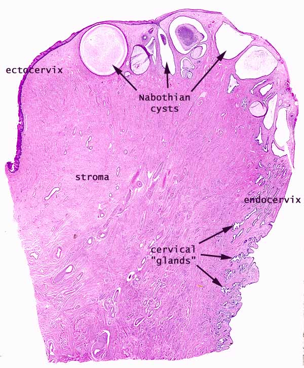
nabothian cyst
endometrial polyp
leiomyoma - fibroid, benign tumor (smooth muscle cells)
Kaposi's sarcoma
appendix
white fibroid? = exudate
fecalith
ovarian cancer
chorioplexus?
teratoma
endometriosis
gliosis
histiocyte
------------------------------------------------------------------------------
Peyer's patches

-------------------------------------------------------------------------------
cryptits
 | ||
| ||
| Microscopy showing the colonic lining with acute inflammatory cells invading into the crypt lining (cryptitis) and the lumen forming a crypt abscess. The surrounding lamina propria shows diffuse and dense infiltration by acute and chronic inflammatory cells. (H/E, 10x) |


The Term mycosis (plural: mycoses) refers to conditions in which fungi pass the resistance barriers of the human or animal body and establish infections. Mycoses are classified according to the tissue levels initially colonized:
- Superficial mycoses - limited to the outermost layers of the skin and hair.
- Cutaneous mycoses - extend deeper into the epidermis, as well as invasive hair and nail diseases. These diseases are restricted to the keratinized layers of the skin, hair, and nails. Unlike the superficial mycoses, host immune responses may be evoked, resulting in pathologic changes expressed in the deeper layers of the skin. The organisms that cause these diseases are called dermatophytes. The resulting diseases are often called ringworm (even though there is no worm involved) or tinea. Cutaenous mycoses are caused by Microsporum, Trichophyton, and Epidermophyton fungi, which together comprise 41 species.
- Subcutaneous mycoses - involve the dermis, subcutaneous tissues, muscle, and fascia. These infections are chronic and can be initiated by piercing trauma to the skin, which allows the fungi to enter. These infections are difficult to treat and may require surgical interventions such as debridement.
- Systemic mycoses due to primary pathogens - originate primarily in the lungs and may spread to many organ systems. Organisms that cause systemic mycoses are inherently virulent. Generally, primary pathogens that cause systemic mycoses are dimorphic.
- Systemic mycoses due to opportunistic pathogens - infections of patients with immune deficiencies who would otherwise not be infected. Examples of immunocompromised conditions include AIDS, alteration of normal flora by antibiotics, immunosuppressive therapy, and metastatic cancer. Examples of opportunistic mycoses include Candidiasis, Cryptococcosis and Aspergillosis.
[edit] Treatment
Clotrimazol is a common agent for fungal infections of the skin, both in humans and animals. It is commonly available both as a cream, and in ear drops
Undecylenic acid (Castor oil derivative) is an effective fungicide for mycotic (fungal) infections.
-------------------------------------------------------------------------------tubular adenoma

keratin pearl

 This is a so-called keratin pearl. In this case, the malignant cells think the center of this little ball of cells is the surface of the skin, and they are busy growing and leaving their keratin in the middle of it.
 -------------------------------------------------------------------------------
pilosebaceous apparatus


Figure 59: Micrographs of sebaceous glands. Left photo shows the relationship between the arrector pili muscle (M) to the hair follicle (F) and sebaceous gland (G). Right photo is a higher magnification of the sebaceous gland.
--------------------------------------------------------------------------------------------------------------
Prostatic intraepithelial neoplasia (PIN) is a precancerous lesion in the prostate gland which is a precursor of [prostate cancer]. Microscopically, PIN is a collection of irregular cells which are fully contained within the gland structure and have not spread to the surrounding tissue. This may be proven by the identification of basal cells forming the supporting layer of the gland. PIN does not require specific therapy, but close follow-up with additional biopsies is warranted. PIN may disappear, remain unchanged, or progress to prostate cancer, often over as many as ten years.
-------------------------------------------------------------------------------Atypia is a clinical term for abnormality in a cell. The term is medical jargon for an atypical cell. It may or may not be a precancerous indication associated with later malignancy, but the level of appropriate concern is highly dependent on the context with which it is diagnosed.
Atypia can be caused by an infection or irritation if diagnosed in a Pap smear, for example. In the uterus it is more likely to be precancerous.
The term atypia is also used dermatoligically and can be a precursor to melanoma.
A dermatological pathology report may show normal (junctional, compound, or intradermal) nevi, various levels of atypia (slight, moderate, severe), or melanoma. Atypia in this context is a precursor to melanoma, but is not yet melanoma.
If a mole shows slight or moderate atypia and margins are clear, no further treatment is typically needed. It would be wise to re-examine if pigmentation recurs after excision. If a mole shows slight or moderate atypia and margins are not clear, it is typical to re-excise or re-shave to get around the lesion.
If a mole shows marked or severe atypia or any degree of pathologist's concern for melanoma, it would be wise to seek professionals for further evaluation.
------------------------------------------------------------------------------A nabothian cyst is a mucus-filled cyst on the surface of the cervix. They are most often caused when new tissue growth blocks the nabothian glands of the cervix; this traps mucosal secretions in small (usually 2-10 mm in diameter) subdermal pockets.
Nabothian cysts appear most often as firm bumps on the cervix's surface. A woman may notice the cyst when inserting a diaphragm or cervical cap, or when doing the cervix check as part of fertility awareness.[1] A gynecologist may notice the cysts during a pelvic exam.
Nabothian cysts are considered harmless and usually disappear on their own. Some women notice they appear and disappear in relation to their menstrual cycle. If a woman is not sure the anomaly she has found on her cervix is a nabothian cyst, a visit to a doctor is recommended to rule out other conditions.[1]
Rarely, nabothian cysts have a correlation with chronic cervicitis, an inflammatory infection of the cervix.
Nabothian cysts are not considered problematic unless they grow very large and present secondary symptoms. A gynecologist may wish to perform a colposcopy or biopsy on a nabothian cyst to check for cancer or other problems. Two methods for removing these cysts include electrocautery and cryofreezing.
Nabothian cysts are also known as nabothian follicles, mucinous retention cysts, or epithelial inclusion cysts.
------------------------------------------------------------------------------
endometrial polyp


------------------------------------------------------------------------------
Kaposi's sarcoma (KS) is a tumor caused by Kaposi's sarcoma-associated herpesvirus (KSHV), also known as human herpesvirus 8 (HHV8). Despite its name, it is generally not considered a true sarcoma, which is a tumor arising from connective tissue. KS actually arises as a cancer of lymphatic endothelium and forms vascular channels that fill with blood cells, giving the tumor its characteristic bruise-like appearance. KS was historically very rare and found mainly in older men of Mediterranean, Jewish or African origin[1] (classic KS), or patients with severely weakened immune systems, such as after an organ transplant (immunosuppressive treatment related KS). In the early 1980s KS began to be seen in AIDS patients. This led to the belief that AIDS weakened the immune system. Kaposi's sarcoma-associated herpesvirus is responsible for all forms of KS.
Symptoms
KS lesions are nodules or blotches that may be red, purple, brown, or black, usually painless but sometimes painful and swollen. They most often appear under the surface of the skin or on mucous membranes, where they are particularly dangerous if they cause enough swelling to obstruct circulation, breathing, or eating. They may also be found in internal organs, particularly the respiratory system or gastrointestinal system; internal lesions are most commonly seen in epidemic KS, and can cause fatal bleeding.
KS can occur among transplant patients, in whom the tumor can disseminate. Stopping immunosuppression can eliminate KS but also can cause rejection of the transplanted organ.
[edit] Pathophysiology and diagnosis
KS lesions contain tumor cells with a characteristic abnormal elongated shape, called spindle cells. The tumor is highly vascular, containing abnormally dense and irregular blood vessels, which leak red blood cells into the surrounding tissue and give the tumor its dark color. Inflammation around the tumor may produce swelling and pain.
Although KS may be suspected from the appearance of lesions and the patient's risk factors, a definite diagnosis can only be made by biopsy and microscopic examination, which will show the presence of spindle cells. Detection of the viral protein LANA in tumor cells confirms the diagnosis.
[edit] Treatment and prevention
Kaposi's sarcoma is not curable, in the usual sense of the word, but it can often be effectively palliated for many years and this is the aim of treatment. In KS associated with immunodeficiency or immunosuppression, treating the cause of the immune system dysfunction can slow or stop the progression of KS. In 40% or more of patients with AIDS-associated Kaposi's sarcoma, the Kaposi lesions will shrink upon first starting highly active antiretroviral therapy (HAART). However, in a certain percentage of such patients, Kaposi's sarcoma may again grow after a number of years on HAART, especially if HIV is not completely suppressed. Patients with a few local lesions can often be treated with local measures such as radiation therapy or cryotherapy. Surgery is generally not recommended as Kaposi's sarcoma can appear in wound edges. More widespread disease, or disease affecting internal organs, is generally treated with systemic therapy with interferon alpha, liposomal anthracyclines (such as Doxil) or paclitaxel.
With the decrease in death rate among AIDS patients receiving new treatments in the 1990s, the incidence and severity of epidemic KS also decreased. However, the number of patients living with AIDS is increasing substantially in the United States, and it is possible that the number of patients with AIDS-associated Kaposi's sarcoma will again rise as these patients live longer with HIV infecton.
Blood tests to detect antibodies against KSHV have been developed and can be used to determine if a patient is at risk for transmitting infection to his or her sexual partner, or if an organ is infected prior to transplantation.
------------------------------------------------------------------------------A fecaloma (meaning a tumor made of feces, also called fecalith and coprolith, i.e., stones made of feces) is a hardening of feces into stones of varying size inside the colon, which may appear whenever chronic obstruction of transit occurs, such as in megacolon and chronic constipation. Some diseases, such as Chagas disease, Hirschsprung's disease and others provoke the destruction of the autonomic nervous system inside the colon's mucosa (Auerbach's plexus) and may cause extremely large (giant) fecalomas, which must be surgically removed (disimpaction). Normally, however, fecalomas can be manually disimpacted or by passing colonic tubes (catheters which carry a flow of disimpaction fluid (solvent).
Fecal impaction may have severe and even lethal effects, such as the rupture of the colon's walls by acute angles of the fecalomas (stercoral perforation), followed by septicemia.
-------------------------------------------------------------------------------A teratoma is a type of neoplasm (specifically, a tumor). The word teratoma comes from Greek and means roughly monstrous tumor. Definitive diagnosis of a teratoma is based on its histology: a teratoma is a tumor with tissue or organ components resembling normal derivatives of all three germ layers. Rarely, not all three germ layers are identifiable. The tissues of a teratoma, although normal in themselves, may be quite different from surrounding tissues, and may be highly inappropriate, even grotesque (hence the monstrous): teratomas (the plural infrequently given as teratomata) have been reported to contain hair, teeth, bone and very rarely more complex organs such as eyeball, torso, and hand. Usually, however, a teratoma will contain no organs but rather one or more tissues normally found in organs such as the brain, liver, and lung.
Teratomas belong to a class of tumors known as nonseminomatous germ cell tumors (NSGCT), all of which are the result of abnormal development of pluripotent (or totipotent) cells: germ cells and embryonal cells. Teratomas of embryonal origin are congenital; teratomas of germ cell origin may or may not be congenital. The kind of pluripotent cell appears to be unimportant, apart from constraining the location of the resulting tumor in the body.
Location
Teratomas derived from germ cells occur in the testes in males and ovaries in females. Teratomas derived from embryonal cells usually occur on the body midline: in the brain, elsewhere inside the skull, in the nose, in the tongue, under the tongue, and in the neck (cervical teratoma), mediastinum, retroperitoneum, and attached to the coccyx. However, teratomas may also occur elsewhere: very rarely in solid organs (most notably the heart and liver) and more commonly on the skull sutures. Embryonal teratomas most commonly occur in the sacrococcygeal region and sacrococcygeal teratoma is the single most common tumor found in newborn babies.
Of teratomas on the skull sutures, approximately 50% are found in or adjacent to the orbit [1].
[edit] Pathology classification of individual teratomas
Teratomas commonly are classified using a grading system: 0 or mature (benign); 1 or immature, probably benign; 2 or immature, possibly malignant (cancerous); and 3 or frankly malignant. See also cancer staging. Teratomas are also classified by their content: a solid teratoma contains only tissues (perhaps including more complex structures); a cystic teratoma contain only pockets of fluid or semi-fluid such as cerebrospinal fluid, sebum, or fat; a mixed teratoma contains both solid and cystic parts. Cystic teratomas usually are grade 0 and, conversely, grade 0 teratomas usually are cystic.
Grade 1 and 2 teratomas have the potential to become malignant (grade 3), and malignant teratomas have the potential to metastasize.
Initial diagnosis
Teratomas are believed to be present since birth, or even before birth, and can therefore be considered congenital tumors. However, many teratomas are not diagnosed until later in childhood or in adulthood. Large tumors are more likely to be diagnosed early on. Sacrococcygeal and cervical teratomas are often detected by prenatal ultrasound. Additional diagnostic methods may include MRI. In rare circumstances, the tumor is so large that the fetus may suffer. In the case of large sacrococcygeal teratomas, a significant portion of the fetus' blood flow is redirected toward the tumor (a phenomenon called steal syndrome), causing heart failure, or hydrops, of the fetus. In selected cases, a fetal operation may be indicated.
Beyond the newborn period, symptoms of a teratoma depend on its location and organ of origin. Ovarian teratomas often present with abdominal or pelvic pain, caused by torsion of the ovary or irritation of its ligaments. Testicular teratomas present as a palpable mass in the testis; mediastinal teratomas often cause compression of the lungs or the airways and may present with chest pain and/or respiratory symptoms.
Some teratomas contain yolk sac elements, which secrete alpha-fetoprotein (AFP). Detection of AFP may help to confirm the diagnosis and is often used as a marker for recurrence or treatment efficacy, but is rarely the method of initial diagnosis. (Maternal serum alpha-fetoprotein, or MSAFP, is a useful screening test for other fetal conditions, including Down syndrome, spina bifida and abdominal wall defects such as gastroschisis).
--------------------------------------------------------------------------------Endometriosis is a common medical condition affecting an estimated 89 million women of reproductive age around the world.[citation needed] In endometriosis, the tissue which lines the uterus (the endometrium, from endo, "inside", and metra, "womb") is found growing outside the uterus, within other areas of the body. Normally, the endometrium is shed each month during the menstrual cycle; however, in endometriosis, the misplaced endometrium is usually unable to exit the body. The endometriotic tissues still detach and bleed, but the result is far different: internal bleeding, degenerated blood and tissue shedding, inflammation of the surrounding areas, and formation of scar tissue may result. In addition, depending on the location of the growths, interference with the normal function of the bowel, bladder, small intestines and other organs within the pelvic cavity can occur. In very rare cases, endometriosis has also been found in the skin, the lungs, the diaphragm, and even the brain.[citation needed]
--------------------------------------------------------------------------------------------------------------
Gliosis is a proliferation of astrocytes in damaged areas of the central nervous system (CNS). Astrocytes are relatively large glial cells and are the connective tissue cells of the CNS. Astrocytes have various functions, including accumulating in areas where nerve cells (neurons) have been damaged. Gliosis and neuronal loss in certain brain regions are findings seen in various neurodegenerative disorders such as Korsakoff's syndrome and AIDS dementia complex.
-------------------------------------------------------------------------------
A histiocyte is a cell that is part of the human immune system. All categories of Histiocytes are derived from the bone marrow by multiplication from a stem cell. The derived cells migrate from the bone marrow to the blood as monocytes. They circulate through the body and stop in various organs where they undergo differentiation into histiocytes which are part of the mononuclear phagocytic system (MPS).
