clinical pathology - lab data, DNA test
margin - surgical edge
FNA(fine needle aspiration) - mostly for cystic mass
- tests for malignancy
- no need for spray
- not for melanoma
cone biopsy?
quick freeze cut
Pap smear - looking for abnormal? dysplastic? => biopsy if +
raisonoid(wrinkled nuclei)
HPV infection precursor to malignancy => cervical carcinoma

Blood bank replace plasma, red cell. Every hospital supposed to have one.
Surgical Pathology
Organ taken out, measure size, photograph, collect lymph nodes, put'em on cassettes, infuse paraffin wax into it. 8 microns thick slices. Put'em on slides for observation.
medical autopsy
check for evidence of pneumonia, heart attack?
Fibroids - can stimulate stroma to secrete estrogen(exceptional case)
Leiomyoma - benign smooth muscle tumor => leiomyosarcoma
uterus smooth muscle, not voluntary - like esophagus, stomach, GI, etc.
syncitiotrophoblast - multiple cells fuse into one
cytotrophoblast
lifelink - for organ transplant
acute tubular necrosis - mudy brown cells(RBC + WBC + protein)
--------------------------------------------------------------------------
In Medicine, the tissue margins are the edges of an excision (or resection) surgical specimen. The margins are examined by a pathologist to evaluate the likelihood that the disease (for example, a cancer) has been completely removed. "Clear margins," or "negative margins", means that the surgeon's cut was in healthy tissue, while "positive margins" means that the surgeon has cut through diseased tissue. This increases the likelihood that some disease was left inside the patient and, therefore, that the disease will recur.
--------------------------------------------------------------------------
In gynecology, the Papanikolaou test or Papanicolaou test (also called Pap smear, Pap test, cervical smear, or smear test) is a medical screening method for detecting infectious, premalignant, and malignant processes in the ectocervix, endocervix and endometrium. The pre-cancerous changes (called dysplasias or cervical or endocervical intraepithelial neoplasia) are usually caused by sexually transmitted human papillomaviruses (HPVs). The test aims to detect and prevent the progression of HPV-induced cervical cancer and other abnormalities in the female genital tract by sampling cells from the outer opening of the cervix (Latin for "neck") of the uterus and the endocervix. The sampling technique changed very little since its invention by Georgios Papanikolaou (1883–1962) to detect cyclic hormonal changes in vaginal cells in the early 20th century until the development of liquid based cell monolayer technology. The test remains a highly effective, widely used method for early detection of cervical cancer and pre-cancer.
It is generally recommended that sexually active females seek Pap smear testing annually, although guidelines may vary from country to country. If results are abnormal, and depending on the nature of the abnormality, the test may need to be repeated in three to twelve months. If the abnormality requires closer scrutiny, the patient may be referred for detailed inspection of the cervix by colposcopy. The patient may also be referred for HPV DNA testing, which can serve as an adjunct (or even as an alternative) to Pap testing.
About 5% to 7% of pap smears produce abnormal results, such as dysplasia, possibly indicating a pre-cancerous condition. Although many low grade cervical dysplasias spontaneously regress without ever leading to cervical cancer, dysplasia can serve as an indication that increased vigilance is needed. Endocervical and endometrial abnormalities can also be detected, as can a number of infectious processes, including yeast and trichomonas. A small proportion of abnormalities are reported as of "uncertain significance".
Technical aspects
Samples are collected from the outer opening or os of the cervix using an Aylesbury spatula or (more frequently with the advent of liquid-based cytology) a plastic-fronded broom. The cells are placed on a glass slide and checked for abnormalities in the laboratory.
Since the mid-1990s, techniques based around placing the sample into a vial containing a liquid medium which preserves the cells have been increasingly used. The media are primarily ethanol based. Two of the types are Sure-Path (TriPath Imaging) and Thin-Prep (Cytyc Corp). Once placed into the vial, the sample is processed at the laboratory into a cell monolayer, stained, and examined by light microscopy. The liquid sample is also suitable for low and high risk HPV testing. Proper sample acquisition is crucial to the accuracy of the test; clearly, a cell that is not in the sample cannot be evaluated. Even liquid based pap smears have a false negative rate of 15-35% and for that reason ACOG and ASCCP have recommended the use of HPV testing in addition to the pap smear in all women over the age of 30. By adding the more sensitive HPV test, which identifies the HPV virus itself, the sensitivity of the pap smear is improved to nearly 100%.
In the last decade there have been successful attempts to develop automated, computer image analysis systems for screening. One of these has been FDA approved and functions in high volume reference laboratories, with human oversight.
The sample is stained using the Papanicolaou technique, in which tinctorial dyes and acids are selectively retained by cells. Unstained cells can not be visualized with light microscopy. The stains chosen by Papanicolau were selected to highlight cytoplasmic keratinization, which actually has almost nothing to do with the nuclear features used to make diagnoses now.
The sample is then screened by a specially trained and qualified cytotechnologist using a light microscope. The terminology for who screens the sample varies according the country; in the UK, the personnel are known as Cytoscreeners, Biomedical scientists (BMS), Advanced Practitioners and Pathologists. The latter two take responsibility for reporting the abnormal sample which may require further investigation.
In the United States, physicians who fail to diagnose cervical cancer from a pap smear have been convicted of negligent homicide. In 1988 and 1989, Karen Smith had received pap smears which were argued to have "unequivocally" shown that she had cancer; yet the lab had not made the diagnosis. She died on March 8, 1995. Later, a physician and a laboratory technician were convicted of negligent homicide. These events have led to even more rigorous quality assurance programs, and to emphasizing that this is a screening, not a diagnostic, test, associated with a small irreducible error rate.
[edit] Practical aspects
The physician or operator collecting a sample for the test inserts a speculum into the patient's vagina, to obtain a cell sample from the cervix. A pap smear appointment is normally not scheduled during menstruation, and patients should avoid contamination of the vagina and uterus (for example by a douche) before the exam, to avoid false results. The procedure is usually just slightly painful, because of the neuroanatomy of the cervix. However, this can depend on the patient's anatomy, the skill of the practitioner, psychological factors, and other conditions. Results usually take about 3 weeks. Slight bleeding, cramps, and other discomfort can occur afterwards.
--------------------------------------------------------------------------Cervical cancer is a malignancy of the cervix. Worldwide, it is the second-most common cancer of women. It may present with vaginal bleeding but symptoms may be absent until the cancer is in its advanced stages, which has made cervical cancer the focus of intense screening efforts utilizing the Pap smear. Most scientific studies have found that human papillomavirus (HPV) infection is responsible for >90% of the cases of cervical cancer. According to a survey of 3,076 women 18 to 75 years of age, awareness about human papillomavirus (HPV) infection and its link to cervical cancer, is relatively low among American women. In 2006, an estimated 10,000 women in the United States will be diagnosed with this type of cancer and nearly 4,000 will die from it.[1] There are 7 most common types of HPV: 16, 18, 31, 33, 42, 52 and 58.[2] Types 16 and 18 are the most common cause of the cancer. There are “low-risk” viruses which does not commonly turn into cancer and “high-risk” viruses that are most likely to develop into cervical cancer; although both can. Having several sexual partners is a major risk factor for developing HPV; although most of the HPV infections clear up on their own, the infections could increase to major abnormalities or cervical cancer.[3] Testing can be done to samples of cervical cells to determine types of HPV that may be present. In some cases, HPV clears up on its own, and in some, there were no signs until it was too late, and cervical cancer developed. It is important to protect yourself from cervical cancer, and a major step in prevention is to get tested for human papillomavirus.
Treatment consists of surgery (including local excision) in early stages and chemotherapy and radiotherapy in advanced stages of the disease. An effective vaccine, the HPV vaccine, for the two most common strains of HPV has recently been licenced (see Vaccine section, below).
--------------------------------------------------------------------------A leiomyoma (plural is 'leiomyomata') is a benign smooth muscle neoplasm that is not premalignant. They can occur in any organ, but the most common forms occur in the uterus and the esophagus.
Etymology

[edit] Uterine leiomyomata
Uterine fibroids are leiomyomata of the uterine smooth muscle. As other leiomyomata, they are benign, but may lead to excessive menstrual bleeding (menorrhagia), often cause anemia and may lead to infertility. Enucleation is removal of fibroids without removing the uterus (hysterectomy), which is also commonly performed. Laser surgery (called myolysis) is increasingly used, and provides a viable alternative to surgery.
Uterine leiomyomas originate in the myometrium and are classified by location:
- Submucous – lie just beneath the endometrium.
- Intramural – lie within the uterine wall.
- Subserous – lies at the serosal surface of the uterus or may bulge out from the myometrium and can become pedunculated.
Estrogen and progesterone usually stimulate their growth, and hormone suppression may hence decrease their size.
[edit] Esophageal
They are also the most common benign esophageal tumour, though this accounts for less than 1% of esophageal neoplasms. The remainder consists mainly of carcinomas
--------------------------------------------------------------------------The cytotrophoblast (or layer of Langhans) is the inner layer of the trophoblast, interior to the syncytiotrophoblast.
Cytotrophoblasts are stem cells in the chorionic villous. During differentiation, mononuclear cytotrophoblast fuse together into the multinucleated syncytiotrophoblasts.
[edit] Invading cytotrophoblasts
These cells invade maternal uterine tissues and thus are in direct contact with maternal stroma and immune cells.
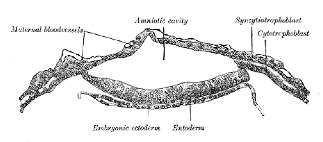
Section through embryonic area of Vespertilio murinus to show the formation of the amniotic cavity.

primary chorionic villi

secondary chorionic villi
--------------------------------------------------------------------------
Chorion
| Chorion | |
|---|---|
 | |
| Diagram showing earliest observed stage of human ovum. | |
 | |
| Human foetus, enclosed in the amnion. | |
| Gray's | subject #12 60 |
| MeSH | Chorion |
The chorion surrounds the embryo and other membranes. It consists of two layers: an outer formed by the primitive ectoderm or trophoblast, and an inner by the somatic mesoderm; with this latter the amnion is in contact.
The trophoblast is made up of an internal layer of cubical or prismatic cells, the cytotrophoblast or layer of Langhans, and an external layer of richly nucleated protoplasm devoid of cell boundaries, the syncytiotrophoblast.
It undergoes rapid proliferation and forms numerous processes, the chorionic villi, which invade and destroy the uterine decidua and at the same time absorb from it nutritive materials for the growth of the embryo.
The chorionic villi are at first small and non-vascular, and consist of trophoblast only, but they increase in size and ramify, while the mesoderm, carrying branches of the umbilical vessels, grows into them, and in this way they are vascularized.
Blood is carried to the villi by the branches of the umbilical arteries, and after circulating through the capillaries of the villi, is returned to the embryo by the umbilical veins. Until about the end of the second month of pregnancy the villi cover the entire chorion, and are almost uniform in size, but after this they develop unequally.
The greater part of the chorion is in contact with the decidua capsularis, and over this portion the villi, with their contained vessels, undergo atrophy, so that by the fourth month scarcely a trace of them is left, and hence this part of the chorion becomes smooth, and is named the chorion læve; as it takes no share in the formation of the placenta, it is also named the non-placental part of the chorion.
On the other hand, the villi on that part of the chorion which is in contact with the decidua placentalis increase greatly in size and complexity, and hence this part is named the chorion frondosum.
--------------------------------------------------------------------------Chorionic villi
The chorion undergoes rapid proliferation and forms numerous processes, the chorionic villi, which invade and destroy the uterine decidua and at the same time absorb from it nutritive materials for the growth of the embryo.
The chorionic villi are at first small and non-vascular, and consist of trophoblast only, but they increase in size and ramify, while the mesoderm, carrying branches of the umbilical vessels, grows into them, and in this way they are vascularized.
Blood is carried to the villi by the branches of the umbilical arteries, and after circulating through the capillaries of the villi, is returned to the embryo by the umbilical veins.
Until about the end of the second month of pregnancy the villi cover the entire chorion, and are almost uniform in size, but after this they develop unequally.
The greater part of the chorion is in contact with the decidua capsularis, and over this portion the villi, with their contained vessels, undergo atrophy, so that by the fourth month scarcely a trace of them is left, and hence this part of the chorion becomes smooth, and is named the chorion læve; as it takes no share in the formation of the placenta, it is also named the non-placental part of the chorion.
On the other hand, the villi on that part of the chorion which is in contact with the decidua placentalis increase greatly in size and complexity, and hence this part is named the chorion frondosum.
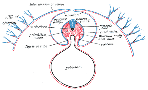
Diagram of a transverse section, showing the mode of formation of the amnion in the chick. The amniotic folds have nearly united in the middle line. Ectoderm, blue; mesoderm, red; entoderm and notochord, black. (Villi of chorion labeled at upper left.)
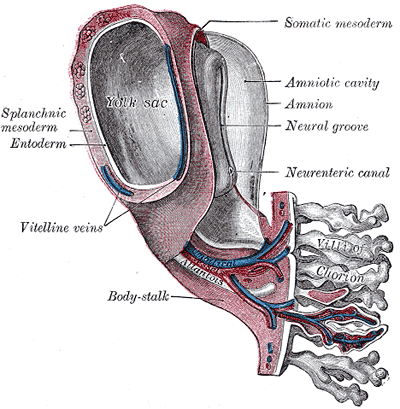
Model of human embryo 1.3 mm. long. (Villi of chorion labeled at lower right.)
Acute tubular necrosis or (ATN) is a medical condition involving the death of tubular cells that form the tubule that transports urine to the ureters while reabsorbing 99% of the water (and highly concentrating the salts and metabolic byproducts). Tubular cells continually replace themselves and if the cause of ATN is removed then recovery is likely. ATN presents with acute renal failure to the point that the two concepts are used interchangeably.
It may be classified as either toxic or ischemic. Toxic ATN occurs when the tubular cells are exposed to a toxic substance (nephrotoxic ATN). Ischemic ATN occurs when the tubular cells do not get enough oxygen, a condition they are highly senistive to due to their very high metabolism.
[edit] Toxic ATN
Toxic ATN can be caused by free hemoglobin or myoglobin, by medication such as antibiotics and cytostatic drugs, or by intoxication (ethylene glycol, "anti-freeze").
Histopathology: Toxic ATN is characterized by proximal tubular epithelium necrosis (no nuclei, intense eosinophilic homogeneous cytoplasm, but preserved shape) due to a toxic substance (poisons, organic solvents, drugs, heavy metals). Necrotic cells fall into the tubule lumen, obliterating it, and determining acute renal failure. Basement membrane is intact, so the tubular epithelium regeneration is possible. Glomeruli are not affected.
[edit] Ischemic ATN
Ischemic ATN can be caused when the kidneys are not sufficiently perfused for a long period of time (i.e. renal artery stenosis) or during shock. Hypoperfusion can also be caused by embolism of the renal arteries. Ischemic ATN specifically causes skip lesions through the tubules.
many nuclei on normal cells
scarce nuclei on necrotic tissue
