lipase, amylase
if elevated, => biliary problem
pancreatitis
alkaline phosphate up, ancillary glucose up
Aspiration vs. endoscopic intervention
unnecessary ERCP(endoscopic retrograde cholangiopancreatography), unsuspected common gallstone, sphincterotomy
risks => pancreatic, duodenal perforation, fistula, bleeding
neuroendocrine tumor(pancreatic, for palliative resection)
RTC - return to clinic
IDC, DCIS - invasive ductal carcinoma, ductal carcinoma in situ
3+ for estrogen receptor, PR(-)
FNA - fine needle aspiration
axillary nodal resection - palpable or not palpable?
tru-cut needle biopsy vs. FNA
axillary lipoma
ancillary glucose up
--------------------------------------------------------------------------
gastric adenocarcinoma
GE Junction mass
M > F, B > W
risk factors dietary
- high carb
- low animal protein
- high salt meat/fish (lack of refrigeration)
- high nirate
- low raw vegetable, fresh fruit, high fiber bread
- low SES(socioeconomic status)
- smoking
- H. pylori
- pernicious anemia history
- gastric polyp
- gastritis, gastric atrophy
Pathology
- 95% adenocarcinoma
- Borrman classification(protrude type / depressed types (3))
- linitis plastica: type 4 involving entire stomach, leather bottle in Latin?
clinical manifestation
- nonspecific early symptoms, vague epigastric pain
- advanced stages: anorexic, weight loss, nausea, hematemesis, hepatomegaly, jaundice, ascites
4 Signs of gastric tumor 1) Virschow's node 2) Sister Mary Joseph's node 3) Blumer's Shelf 4) Krunkenberg tumor
Pouch of Douglas - pouch between rectum and bladder/uterus
Diagnosis
- upper GI biopsy
- CXR, CT, A/P, CT chest barium proximal lesion
- laparoscopy to detect metastatic
TMN staging
T (primary tumor), N (regional lymph node), M (distant metastasis), R status (tumor after resection)
nodes for gastric cancer depends on part of stomach
treatment
- resection line at least 6 cm from tumor mass
- proximal lesions - total gastrectomy(big margin to clear submucosal extension) / proximal gastric resection
- distal lesions - subtotal gastrectomy
- role of extended lymphadenectomy controversial
don't freeze margin till higher up
sudden passage of food content to duodenum by taking out pylorus
vagatomy - why lead to dumping?
pylorus/antrum - empty solid part of food
body/proximal stomach empty liquid
vagus nerve controls internal sphincter, pylorus, etc.
If vagatomy, then internal sphincter relaxes and remains open
pylorus stays closed
gotta do something that allow pylorus to empty
early gastric cancer = not a clinical disease but a pathologic disease
treatment
adjuvant therapy - 5FU, leucovorin
palliation - dilation / stent placement
1) try to cure the pt early
2) palliate the pt if late - death from gastric cancer miserable
why colon cancer better prognosis than pancreatic / gastric tumor?
- tumor in stomach, due to the size of stomach, is able to grow large till symptomatic
- pancreatic tumor, due to its location, can also grow large till symptomatic
- frequency of colon cancer high => screen early => pick up
- gastric cancer infrequent, less screening
follow up
- recurrence in 3 yrs
lymph node metastasis
distal gastric tumor - spread to lymph nodes of duodenum
proximal gastric tumor - spread to spleen lymph nodes
--------------------------------------------------------------------------
Pancreatic juice is composed of two secretory products critical to proper digestion: digestive enzymes and bicarbonate. The enzymes are synthesized and secreted from the exocrine acinar cells, whereas bicarbonate is secreted from the epithelial cells lining small pancreatic ducts.
Digestive Enzymes
The pancreas secretes a magnificent battery of enzymes that collectively have the capacity to reduce virtually all digestible macromolecules into forms that are capable of, or nearly capable of being absorbed. Three major groups of enzymes are critical to efficient digestion:
1. Proteases
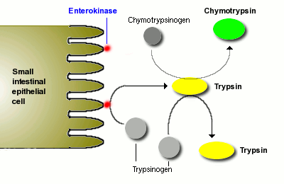
Digestion of proteins is initiated by pepsin in the stomach, but the bulk of protein digestion is due to the pancreatic proteases. Several proteases are synthesized in the pancreas and secreted into the lumen of the small intestine. The two major pancreatic proteases are trypsin and chymotrypsin, which are synthesized and packaged into secretory vesicles as an the inactive proenzymes trypsinogen and chymotrypsinogen.
As you might anticipate, proteases are rather dangerous enzymes to have in cells, and packaging of an inactive precursor is a way for the cells to safely handle these enzymes. The secretory vesicles also contain a trypsin inhibitor which serves as an additional safeguard should some of the trypsinogen be activated to trypsin; following exocytosis this inhibitor is diluted out and becomes ineffective - the pin is out of the grenade.
Once trypsinogen and chymotrypsinogen are released into the lumen of the small intestine, they must be converted into their active forms in order to digest proteins. Trypsinogen is activated by the enzyme enterokinase, which is embedded in the intestinal mucosa.
Once trypsin is formed it activates chymotrypsinogen, as well as additional molecules of trypsinogen. The net result is a rather explosive appearance of active protease once the pancreatic secretions reach the small intestine.
Trypsin and chymotrypsin digest proteins into peptides and peptides into smaller peptides, but they cannot digest proteins and peptides to single amino acids. Some of the other proteases from the pancreas, for instance carboxypeptidase, have that ability, but the final digestion of peptides into amino acids is largely the effect of peptidases on the surface of small intestinal epithelial cells. More on this later.
2. Pancreatic Lipase
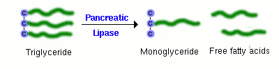
A major component of dietary fat is triglyceride, or neutral lipid. A triglyceride molecule cannot be directly absorbed across the intestinal mucosa. Rather, it must first be digested into a 2-monoglyceride and two free fatty acids. The enzyme that performs this hydrolysis is pancreatic lipase, which is delivered into the lumen of the gut as a constituent of pancreatic juice.
Sufficient quantities of bile salts must also be present in the lumen of the intestine in order for lipase to efficiently digest dietary triglyceride and for the resulting fatty acids and monoglyceride to be absorbed. This means that normal digestion and absorption of dietary fat is critically dependent on secretions from both the pancreas and liver.
Pancreatic lipase has recently been in the limelight as a target for management of obesity. The drug orlistat (Xenical) is a pancreatic lipase inhibitor that interferes with digestion of triglyceride and thereby reduces absorption of dietary fat. Clinical trials support the contention that inhibiting lipase can lead to significant reductions in body weight in some patients.
3. Amylase
The major dietary carbohydrate for many species is starch, a storage form of glucose in plants. Amylase (technically alpha-amylase) is the enzyme that hydrolyses starch to maltose (a glucose-glucose disaccharide), as well as the trisaccharide maltotriose and small branchpoints fragments called limit dextrins. The major source of amylase in all species is pancreatic secretions, although amylase is also present in saliva of some animals, including humans.
Other Pancreatic Enzymes
In addition to the proteases, lipase and amylase, the pancreas produces a host of other digestive enzymes, including ribonuclease, deoxyribonuclease, gelatinase and elastase.
Bicarbonate and Water
Epithelial cells in pancreatic ducts are the source of the bicarbonate and water secreted by the pancreas. Bicarbonate is a base and critical to neutralizing the acid coming into the small intestine from the stomach. The mechanism underlying bicarbonate secretion is essentially the same as for acid secretion parietal cells and is dependent on the enzyme carbonic anhydrase. In pancreatic duct cells, the bicarbonate is secreted into the lumen of the duct and hence into pancreatic juice.
--------------------------------------------------------------------------Alkaline phosphatase (ALP) (EC 3.1.3.1) is a hydrolase enzyme responsible for removing phosphate groups in the 5- and 3- positions from many types of molecules, including nucleotides, proteins, and alkaloids. The process of removing the phosphate group is called dephosphorylation. As the name suggests, alkaline phosphatases are most effective in an alkaline environment.
Physiology
In humans, alkaline phosphatase is present in all tissues throughout the entire body, but is particularly concentrated in liver, bile duct, kidney, bone, and the placenta. The optimal pH for the enzyme activity is pH=10 in standard conditions (310K,1 atm)
[edit] Diagnostic use
Concentrations blood plasma (serum) levels of ALP are typically 30-150 Units per liter, depending on the assay and local normal guidelines.
Lowered levels of ALP are less common than elevated levels.
The following conditions can cause abnormal levels of ALP:
[edit] Elevated levels (hyperphosphatasemia)
If it is unclear why alkaline phosphatase is elevated, isoenzyme studies using electrophoresis can confirm the source of the ALP. Heat stability also distinguishes bone and liver isoenzymes ("bone burns, liver lasts").
- Liver (Liver ALP):
- Bone disease (Bone ALP):
- Paget's disease, osteosarcoma, bone metastases of prostatic cancer (High / very high ALP values)
- Other bone metastases
- Fractured bone
- Multiple myeloma (only when associated with fractures)
- Skeletal involvement of other primary diseases:
- Osteomalacia, rickets, vitamin D deficiency, (Moderate rise)
- Malign tumors (ALP originating from tumor)
- Renal disease (secondary hyperparathyroidism)
- Primary hypothyroidism
- Polycythemia vera
- Myelofibrosis
- Leukemoid reaction to infection
- women on oral contraceptives
- Pregnancy
- Biliary obstruction
- Transient hyperphosphatasaemia of infancy: benign, often assocoated with infection
[edit] Lowered levels (hypophosphatasemia)
- Hypophosphatasia, an autosomal recessive disease
- Postmenopausal women receiving estrogen therapy because of osteoporosis
- Men with recent heart surgery, malnutrition, magnesium deficiency, hypothyroidism or severe anemia
- Children with achondroplasia and cretinism
- Children after a severe enteritis
- Pernicious anemia
- Aplastic anemia
- Chronic myelogenous leukemia
Main Entry: 1an·cil·lary
Pronunciation:
 an(t)-s
 -
- ler-
ler- , esp British an-sil--r
Function: adjective
: being auxiliary or supplementary
Indications
ERCP may be performed for diagnostic or therapeutic reasons.
[edit] Diagnostic
- Obstructive jaundice - This may be due to several causes
- Chronic pancreatitis - a now controversial indication due to widespread availability of safer diagnostic modalities including endoscpoic ultrasound, high-resolution CT, and MRI/MRCP
- Gallstones with dilated bile ducts on ultrasonography
- Bile duct tumors
- Suspected injury to bile ducts either as a result of trauma or iatrogenic
- Sphincter of Oddi dysfunction
- Pancreatic tumors no longer represent a valid diagnostic indication for ERCP unless they cause bile duct obstruction and jaundice. Endoscopic ultrasound represents a safer and more accurate diagnostic alternative
[edit] Therapeutic
- Any of the above when the following may become necessary
- Endoscopic sphincterotomy (both of the biliary and the pancreatic sphincters)
- Removal of stones
- Insertion of stent(s)
- Dilation of strictures (e.g. primary sclerosing cholangitis, anastomotic strictures after liver transplantation)
• Sphincterotomy (sfingk-te-ROT-ohme).
If x-rays show a gallstone or other
blockage, your doctor may choose to
treat it by enlarging the opening of your
bile duct. He or she will make a small cut
in the bile duct with an electrically heated
wire. You will not feel this cut. Gallstones
will be crushed through use of a special
instrument. The tiny pieces will either be
removed using a balloon, or they will be
left to pass into your intestines, where
they will be eliminated naturally.
• Stenting. If your doctor notices a narrowed
duct, he or she may choose to place
a stent in the duct. A stent is a tiny plastic
tube that can help keep a duct open.
Stenting can
relieve jaundice (a condition caused by an
interruption in bile processing) by allowing
bile to drain into the intestines. Stents
are sometimes placed in the pancreatic
duct when it is blocked. Stents can be
pushed through the endoscope and placed
into a narrowed area of the bile duct.
• Nasobiliary (NAY-zoh-BILL-ee-air-ee)
draining. Sometimes, a long, thin plastic
tube, called a nasobiliary tube, is left in
the bile duct and brought out through the
nose. This allows bile to drain so more
x-rays can be taken. The tube may be
kept in for a few days. It may be slightly
uncomfortable at first, but it will not interfere
with eating or drinking.
--------------------------------------------------------------------------
Neuroendocrine tumors, or more properly gastro-entero-pancreatic or gastroenteropancreatic neuroendocrine tumors (GEP-NETs), are cancers of the interface between the endocrine (hormonal) system and the nervous system.
Summary of human GEP-NETs by site of origin and by symptom
- carcinoids (about two thirds of GEP-NETs)
- with carcinoid syndrome (about 10 percent of carcinoids)
- without carcinoid syndrome (about 90 percent of carcinoids)
- PETs (about one third of GEP-NETs)
- nonfunctioning (15 to 30 percent of PETs)
- functioning (70 to 85 percent of PETs)
- gastrinoma, producing excessive gastrin and causing Zollinger-Ellison Syndrome (ZES)
- insulinoma, producing excessive insulin
- glucagonoma, producing excessive glucagon
- vasoactive intestinal peptideoma (VIPoma), producing excessive vasoactive intestinal peptide (VIP)
- PPoma, producing excessive pancreatic polypeptide (often classed with nonfunctioning PETs)
- somatostatinoma, producing excessive somatostatin
- watery diarrhea, hypokalemia-achlorhydria (WDHA)
- CRHoma, producing excessive corticotropin-releasing hormonse (CRH)
- calcitoninoma, producing excessive calcitonin
- GHRHoma, producing excessive growth-hormone-releasing hormone (GHRH)
- neurotensinoma, producing excessive neurotensin
- ACTHoma, producing excessive adrenocorticotropic hormone (ACTH)
- GRFoma, producing excessive growth-hormone release factor (GRF)
- parathyroid hormone–related peptide tumor
- rare GEP-NETs
- medullary carcinoma of the thyroid
- Merkel cell cancer (trabecular cancer)
- small-cell lung cancer (SCLC)
- large-cell neuroendocrine carcinoma (of the lung)
- neuroendocrine carcinoma of the cervix
- Multiple Endocrine Neoplasia type 1 (MEN-1 or MEN1) (usually nonfunctioning) (also causing ZES)
- Multiple Endocrine Neoplasia type 2 (MEN-2 or MEN2)
- neurofibromatosis type 1
- tuberous sclerosis
- von Hippel-Lindau (VHL) disease
- neuroblastoma
- pheochromocytoma (phaeochromocytoma)
- paraganglioma
- neuroendocrine tumor of the anterior pituitary
- Carney's complex
Summary of classification by cell characteristics (the WHO classification)
- Superclass:
- Öberg, WHO, Klöppel et alia: gastro-entero-pancreatic neuroendocrine tumor (GEP-NET)
- Subclass 1 (less malignant)
- Öberg: carcinoid
- WHO: neuroendocrine tumor (NET)
- Klöppel et alia: well-differentiated neuroendocrine tumor (NET) (carcinoid)
- this article: carcinoid
- Subclass 2 (more malignant)
- Öberg: endocrine pancreatic tumor
- WHO: neuroendocrine carcinoma (NEC)
- Klöppel et alia: well-differentiated neuroendocrine carcinoma (NEC) (malignant carcinoid)
- this article: pancreatic endocrine tumor (PET) or endocrine pancreatic tumor (EPT) or islet cell tumor or noncarcinoid GEP-NET
- Subclass 3 (most malignant)
- WHO: poorly-differentiated neuroendocrine carcinoma
- Klöppel et alia: poorly-differentiated neuroendocrine carcinoma (high-grade malignant carcinoid)
- Subclass 4 (mixed)
- WHO: mixed endocrine/exocrine tumor
- Subclass 5 (miscellaneous)
- WHO: rare neuroendocrine-like lesions
GEP-NETs are also sometimes called APUDomas, but that term is now considered to be misleading, since it is based on a discredited theory of the development of the tumors. [8]
Metastases and malignancy
In the context of GEP-NETs, the terms metastatic and malignant are often used interchangeably.
GEP-NETs are often malignant, since the primary site often eludes detection for years, sometimes decades – during which time the tumor has the opportunity to metastasize. Researchers differ widely in their estimates of malignancy rates, especially at the level of the secretory subtypes (the various "-omas").
The most common metastatic sites are the liver, the lymph nodes, and the bones. Liver metastases are so frequent and so well-fed that for many patients, they dominate the course of the cancer. For a patient with a nonsecretory PET, for example, the primary threat to life may be the sheer bulk of the tumor load in the liver.
The term lesion is a synonym for tumor; in particular, lesion means primary tumor, or metastasis.
The list of potential markers for GEP-NETs is long. Aside from the hormones of secretory tumors, the most important markers are
- chromogranin A (CgA)
- urine 5-hydroxy indole acetic acid (5-HIAA) (grade C)
- neuron-specific enolase (NSE, gamma-gamma dimer)
- synaptophysin (P38)
and other markers include
- synaptobrevin (VAMP-1)
- synapsin (1A, 1B, 2A, 2B)
- SV2
- protein P65
- protein S-100
- protein gene product (PGP) 9.5
- intermediate filaments (cytokeratins, vimentin, neurofilaments)
- protein 7B2
- chromogranin B (secretogranin I)
- chromogranin C (secretogranin II)
- pancreastatin
- vasostatin
- cytochrome b561
- leu-7 (HNK-1)
- calcitonin
- human chorionic gonadotropin-alpha (HCG-α)
- human chorionic gonadotropin-beta (HCG-β)
- thyroid function tests (TFTs)
- parathyroid hormone (PTH)
- calcium
- prolactin
- {alpha}-fetoprotein
- carcinoembryonic antigen (CEA)
- ß-human chorionic gonadotrophin (ß-HCG) (grade D)
- CGRP
- GRP
- PYY
- hCGα
- N Peptide K
- neurokinin A
- serotonin
- neurotensin
- motilin
- substance P
- histamine
- catecholamines
- dopa
- various rarer peptide hormones
- synaptotagmin
- HISL-19
and newer (as of 2005) markers include
- N-terminally truncated variant of heat shock protein 70 (Hsp70)
- CDX-2, a homeobox gene product
- neuroendocrine secretory protein-55
Surgery and chemotherapy
Surgery is the only therapy that can cure GEP-NETs. However, the typical delay in diagnosis, giving the tumor the opportunity to metastasize, makes most GEP-NETs ineligible for surgery (non-resectable).
There is "no established standard therapy for the liver metastasis of pancreatic endocrine tumors" (Sato et al. 2000, [16]). The most common nonsurgical therapy for all GEP-NETs is chemotherapy, although chemotherapy is reported to be largely ineffective for carcinoids, not particularly durable (long-lasting) for PETs, and inappropriate for PETs of nonpancreatic origin. [9]
When chemotherapy fails, the most common therapy, in the United States, is more chemotherapy, with a different set of agents. Some studies have shown that the benefit from one agent is not highly predictive of the benefit from another agent, except that the long-term benefit of any agent is likely to be low.
Strong uptake of somatostatin analogs is a negative indication for chemo.
[edit] Symptomatic relief
There are two major somatostatin-analog-based targeted therapies. The first of the two therapies provides symptomatic relief for patients with secretory tumors. In effect, somatostatin given subcutaneously or intramuscularly "clogs up" the receptors, blocking the secretion of hormones from the tumor cells. Thus a patient who might otherwise die from severe diarrhea caused by a secretory tumor can gain additional years of life.
Specific counter-hormones or other hormone-blocking medications are sometimes also used to provide symptomatic relief.
[edit] Hormone-delivered radiotherapy – PRRT
The second of the two major somatostatin-analog-based targeted therapies is called peptide receptor radionuclide therapy (PRRT), though we might simply call it hormone-delivered radiotherapy. In this form of radioisotope therapy (RIT), radioactive substances (called radionuclides or radioligands) are chemically conjugated with hormones (peptides or neuroamines); the combination is given intravenously to a patient who has good uptake of the chosen hormone. The tumor cells attract the hormone, and the attached radiation kills nearby cells. In patients with strongly overexpressing tumor cells, nearly all the radiation either sticks to the tumors or is excreted in urine. As Rufini et alia say, GEP-NETs "are characterized by the presence of neuroamine uptake mechanisms and/or peptide receptors at the cell membrane, and these features constitute the basis of the clinical use of specific radiolabeled ligands, both for imaging and therapy" (Rufini, Calcagni, and Baum 2006, [17]).
The use of PRRT for GEP-NETs is similar to the use of iodine-131 as a standard therapy (in use since 1943) for nonmedullary thyroid tumors (which are not GEP-NETs). Thyroid cells (whether normal or neoplastic) tend to be avid for iodine, and nearby cells are killed when iodine-131 is infused into the bloodstream and is soon attracted to thyroid cells. Similarly, overexpressing GEP-NET cells (neoplastic cells only) are avid for somatostatin analogs, and nearby cells are killed when radionuclides attached to somatostatin analogs are infused into the bloodstream and are soon attracted to the tumor cells. In both therapies, hormonal targeting delivers a much higher dose of radiation than external beam radiation could safely deliver.
As of 2006, PRRT is available in at least dozen medical centers in Europe. In the USA it is FDA-approved, and available at the MD Anderson Cancer Center, but using a radionuclide, indium-111, that is much weaker than the lutetium-177 and the even stronger yttrium-90 used on the European continent. In the UK, only the radionuclide metaiodobenzylguanidine (I-MIBG) is licensed (but GEP-NETs are rarely avid for MIBG). PRRT with lutetium or yttrium is nowhere an "approved" therapy, but the German health insurance system, for example, covers the cost for German citizens.
PRRT using yttrium or lutetium was first applied to humans about 1999. Practitioners continue to refine their choices of radionuclides to maximize damage to tumors, of somatostatin analogs to maximize delivery, of chelators to bind the radionuclides with the hormones (and chelators can also increase uptake), and of protective mechanisms to minimize damage to healthy tissues (especially the kidneys). [10]
[edit] Hepatic artery-delivered therapies
One therapy for liver metastases of GEP-NETs is hepatic artery embolization (HAE). Larry Kvols, of the Moffitt Cancer Center and Research Institute in Tampa, Florida, says that "hepatic artery embolization has been quite successful. During that procedure a catheter is placed in the groin and then threaded up to the hepatic artery that supplies the tumors in the liver. We inject a material called embospheres [tiny spheres of glass or resin, also called microspheres] into the artery and it occludes the blood flow to the tumors, and in more than 80% of patients the tumors will show significant tumor shrinkage" (Kvols 2002, [18]). HAE is based on the observation that tumor cells get nearly all their nutrients from the hepatic artery, while the normal cells of the liver get about 75 percent of their nutrients (and about half of their oxygen) from the portal vein, and thus can survive with the hepatic artery effectively blocked. [11]
Another therapy is hepatic artery chemoinfusion, the injection of chemotherapy agents into the hepatic artery. Compared with systemic chemotherapy, a higher proportion of the chemotherapy agents are (in theory) delivered to the lesions in the liver. [12]
Hepatic artery chemoembolization (HACE), sometimes called transarterial chemoembolization (TACE), combines hepatic artery embolization with hepatic artery chemoinfusion: embospheres bound with chemotherapy agents, injected into the hepatic artery, lodge in downstream capillaries. The spheres not only block blood flow to the lesions, but by halting the chemotherapy agents in the neighborhood of the lesions, they provide a much better targeting leverage than chemoinfusion provides.
Radioactive microsphere therapy (RMT) combines hepatic artery embolization with radiation therapy – microspheres bound with radionuclides, injected into the hepatic artery, lodge (as with HAE and HACE) in downstream capillaries. This therapy is also called selective internal radiation therapy, or SIRT. In contrast with PRRT, the lesions need not overexpress peptide receptors. (But PRRT can attack all lesions in the body, not just liver metastases.) Due to the mechanical targeting, the yttrium-labeled microspheres "are selectively taken up by the tumors, thus preserving normal liver" (Salem et al. 2002, [19]). [13]
[edit] Other therapies
Radiofrequency ablation (RFA) is used when a patient has relatively few metastases. In RFA, a needle is inserted into the center of the lesion and is vibrated at high frequency to generate heat; the tumor cells are killed by cooking.
Cryoablation is similar to RFA; an endothermic substance is injected into the tumors to kill by freezing. Cryoablation has been considerably less successful for GEP-NETs than RFA.
Interferon is sometimes used to treat GEP-NETs; its use was pioneered by Dr. Kjell Öberg at Uppsala. For GEP-NETs, Interferon is often used at low doses and in combination with other agents (especially somatostatin analogs such as octreotide). But some researchers claim that Interferon provides little value aside from symptom control.
As described above, somatostatin analogs have been used for about two decades to alleviate symptoms by blocking the production of hormones from secretory tumors. They are also integral to PRRT. In addition, some doctors claim that, even without radiolabeling, even patients with nonsecretory tumors can benefit from somatostatin analogs, which purportedly can shrink or stabilize GEP-NETs. But some researchers claim that this "cold" octreotide provides little value aside from symptom control.
Finally, therapies based on growth factor inhibitors are in the experimental stage. These inhibitors of epidermal growth factor receptors (EGFRs), of vascular endothelial growth factor receptors (VEGFRs), and of angiopoietin-related growth factor (AGF) include imatinib, sunitinib, temozolide, thalidomide, sorafenib, and panitumumab.
--------------------------------------------------------------------------
 The ductal carcinoma is clearly visible as a dominant mass with an irregular fibrotically stranded boundary, and some of the strands seemingly to extend toward the nipple.
Ductal carcinoma is a very common type of breast cancer in women. It comes in two forms: infiltrating ductal carcinoma (IDC), an invasive cell type; and ductal carcinoma in situ (DCIS), a noninvasive cancer.
DCIS is the most common type of noninvasive breast cancer in women. Ductal carcinoma refers to the development of cancer cells within the milk ducts of the breast. In situ means "in place" and refers to the fact that the cancer has not moved out of the duct and into any surrounding tissue. DCIS can be difficult to detect by physical examination and is usually discovered through a mammogram as very small specs of calcium known as microcalcifications. However, not all microcalcifications indicate the presence of DCIS, which must be confirmed by biopsy. DCIS may be multifocal, and treatment is aimed at excising all of the abnormal duct elements, leaving "clear margins", an area of much debate. After excision treatment often includes local radiation therapy.
IDC, formed in the ducts of breast in the earliest stage, is the most common, most heterogeneous invasive breast cancer cell type. It accounts for 80% of all types of breast cancer. On a mammography, it is usually visualized as a mass with fine spikes radiating from the edges, and small microcalcification may be seen as well. On physical examination, this lump usually feels much harder or firmer than the one with benign breast lesions. On microscopic examination, the cancerous cells invade and replace the surrounding normal tissue inside the breast.
Special histologic subtypes of IDC may vary in prognosis, survival, and recurrence rates: the ones with histology of mucinous, papillary, cribriform, and tubular carcinomas have a better prognosis, longer survival, and lower recurrence rates than those with histology like signet-ring cell carcinoma, carcinoma with sarcomatoid metaplasia, and inflammatory carcinoma.
--------------------------------------------------------------------------
Signs of gastric tumor
Virchow's node - metastatic tumor to left supraclavicular node
Sister Mary Joseph's sign/node - metastatic tumor to umbilical lymph node(s)
Blumer's shelf - metastatic disease to the rectouterine ( pouch of Douglas) or rectovesical pouch creating a "shelf" that is palpable on rectal exam
Krukenberg tumor - metastatic tumor to the ovary
--------------------------------------------------------------------------
The vagus nerve (also called pneumogastric nerve or cranial nerve X) is the tenth of twelve paired cranial nerves, and is the only nerve that starts in the brainstem (within the medulla oblongata) and extends, through the jugular foramen, down below the head, to the abdomen.
The medieval Latin word vagus means literally "wandering" (the words vagrant, vagabond, and vague come from the same root). It is also called the pneumogastric nerve since it innervates both the lungs and the stomach.
Innervation
The vagus nerve supplies motor parasympathetic fibers to all the organs except the suprarenal glands, from the neck down to the second segment of the transverse colon. The vagus also controls a few skeletal muscles, namely:
- Levator veli palatini muscle
- Salpingopharyngeus muscle
- Palatoglossus muscle
- Palatopharyngeus muscle
- Superior, middle and inferior pharyngeal constrictors
- Muscles of the larynx (speech).
This means that the vagus nerve is responsible for such varied tasks as heart rate, gastrointestinal peristalsis, sweating, and quite a few muscle movements in the mouth, including speech (via the recurrent laryngeal nerve) and keeping the larynx open for breathing. It also receives some sensation from the outer ear, via the Auricular branch (also known as Alderman's nerve) and part of the meninges.
[edit] The vagus nerve and the heart
Parasympathetic innervation of the heart is mediated by the vagus nerve. The right vagus innervates the Sinoatrial node. Parasympathetic hyperstimulation predisposes those affected to bradyarrhythmias. The left vagus when hyperstimulated predisposes the heart to Atrioventricular (AV) blocks.
At this location Otto Loewi first proved that nerves secrete substances called neurotransmitters which have effects on receptors in target tissues. Loewi described the substance released by the vagus nerve as vagusstoff, which was later found to be acetylcholine.
The vagus nerve has three associated nuclei, the dorsal motor nucleus, the nucleus ambiguus and the solitary nucleus.
Drugs that inhibit the muscarinic cholinergic receptor (anticholinergics) such as atropine and scopolamine are called vagolytic because they inhibit the action of the vagus nerve on the heart, gastrointestinal tract and other organs. Anticholinergic drugs increase heart rate and are used to treat bradycardia(slow heart rate) and asystole, which is when the heart has no electrical activity. Anticholinergic drugs relax the detrusor muscle and cause constipation which again involves the vagus nerve.
Bulemics and anorexics have high vagal activity which is associated with the arrhythmias seen in these patients.
[edit] Medical treatment involving the vagus nerve
Vagus nerve stimulation (VNS) therapy using a pacemaker-like device implanted in the chest is a treatment used since 1997 to control seizures in epilepsy patients and has recently been approved for treating drug-resistant cases of clinical depression. [1] A convenient, non-invasive VNS device that stimulates an afferant branch of the vagus nerve is also being developed and will soon undergo trials.
A degree of intermittent VNS can be achieved by daily breathing exercises (for example, Pranayama) over a period of several weeks. In some patients, such proactive relaxation exercises have been found to correlate with lower blood pressure and lower heart rate and more stable moods. The valsalva maneuver may activate the vagus nerve and is a 'natural' way to achieve the same effect in some patients. Patients with atrial fibrillation, supraventricular tachycardia and other illnesses may be trained to perform the valsalva maneuver (or find it for themselves).
Vagotomy (cutting of the vagus nerve) is a now-obsolete therapy that was performed for peptic ulcer disease.
[edit] Physical and emotional effects
Activation of the vagus nerve typically leads to a reduction in heart rate, blood pressure, or both. This occurs commonly in the setting of gastrointestinal illness such as viral gastroenteritis or acute cholecystitis, or in response to other stimuli, including carotid sinus massage, Valsalva maneuver, or pain from any cause, particularly having blood drawn. When the circulatory changes are great enough, vasovagal syncope results. Relative dehydration tends to amplify these responses.
Excessive activation of the vagal nerve during emotional stress, which is a parasympathetic overcompensation of a strong sympathetic nervous system response associated with stress, can also cause vasovagal syncope because of a sudden drop in blood pressure and heart rate. Vasovagal syncope affects young children and women more often. It can also lead to temporary loss of bladder control under moments of extreme fear.

