- ethmoid bone, lamina there is cribriform
- cribriform plate of ethmoid roofs nasal cavity
DCIS low grade looks more monotous and has cribriform which is in contrast to adenosis which looks may have fibrosis between glands but does not have cribriform appearance
---------------------------------------------------------
Giemsa stain
- is used for the histopathological diagnosis of malaria and other parasites. It is a mixture of methylene blue and eosin.
- It is specific for the phosphate groups of DNA and attaches itself to regions of DNA where there are high amounts of adenine-thymine bonding. Giemsa stain is used in Giemsa banding, commonly called G-banding, to stain chromosomes and often used to create a karyotype. It can identify chromosomal aberrations such as translocations and interchanges.
- It can be used to study the adherence of pathogenic bacteria to human cells. It differentially stains human and bacterial cells purple and pink respectively. It can be used for histopathological diagnosis of malaria and some other spirochete and protozoan blood parasites.
- Erythrocytes stain pink, platelets show a light pale pink, lymphocyte cytoplasm stains sky blue, monocyte cytoplasm stains pale blue, and leukocyte nuclear chromatin stains magenta.
--------------------------------------------------------
CMV invades vessel and stroma while herpes infects epithelium
Can happen not only in skin but also GI like esophagus
---------------------------------------------------------
BRCA - breast, ovarian and colon
Collagenous spherulosis of the breast is an uncommon localized pattern of basement membrane material deposition that may be mistaken for atypical proliferations or carcinoma.
- stimulte cribriform DCIS
- failure to recognize CS as benign lesion, 28% were misdiagnosed as atypical/malignant
- usually eosinophilic, but this case below has special cases of CS with basophilic spherules
- mucinous early on and progress to collagenous

Figure 1.
 Mucinous/basophilic spherulosis, fibrillary pattern. Radiating basophilic fibrils frequently have a central ringlike structure (hematoxylin-eosin, original magnification ×200).
 Figure 2.Mucinous/basophilic spherulosis, diffuse pattern. This pattern mimics cribriform ductal carcinoma in situ, but a thin, slightly scalloped cuticle is present at the periphery of the spaces (hematoxylin-eosin, original magnification ×320).
Figure 3.Spherulosis with predominantly clear pattern. Spaces with only barely visible material on hematoxylin-eosin staining are separated by rigid eosinophilic septa (hematoxylin-eosin original magnification ×180).
Figure 4.Alcian blue stain shows a pattern similar to that seen in mucinous spherulosis (same case as Figure 3) (Alcian blue, original magnification ×250).
Figure 5.Immunoperoxidase stain for smooth muscle actin on a case of mucinous spherulosis outlines myoepithelial cells located peripherally and around the spherules (original magnification ×450).
Figure 6.Basophilic mucin in ordinary hyperplasia. Note the irregular distribution of secondary lumens and of their basophilic contents (hematoxylin-eosin, original magnification ×275).
---------------------------------------------------------
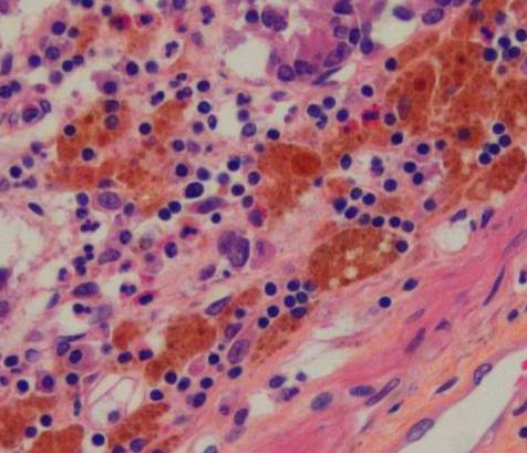
Melanosis coli is a disorder of pigmentation of the wall of the colon, identified at the time of colonoscopy. It is benign, and has no significant correlation with disease. The name is considered a misnomer: the brown pigment seen in the colon is actually lipofuscin, not melanin as the name implies.
the surreptitious use of laxatives, and commonly anthraquinone containing laxatives such as Senna and other plant glycosides. [1] Endoscopically, the mucosa shows a brownish discoloration in a moire pattern. On biopsy, melanosis coli shows characteristic pigment-laden macrophages within the submucosa on PAS staining.[2]
--------------------------------------------------------
Russell Body

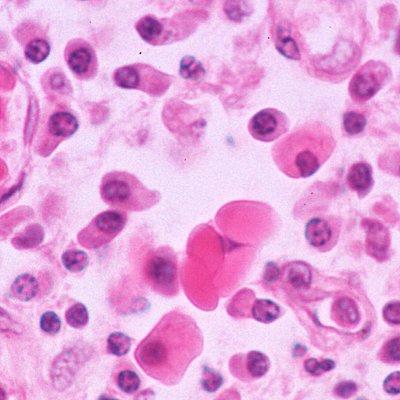
Russell bodies are round accumulations of monoclonal protein that are (or used to be) inside the endoplasmic reticulum, usually immunogloublin within plasma cells. Nobody knows how or why they form, though the question has been addressed by elite pathologists
Plasma cells loaded with Russell bodies are called "Mott cells". These may be so abundant in helicobacter or other gastritis that a pathologist may entertain a more serious diagnosis
The only time that, to my knowledge, Russell bodies make a major difference is in the autopsy confirmation of African sleeping sickness. IgM-based Russell bodies abound in the plasma cells within the brain.
-------------------------------------------------------------------------------
Hep B and C appearance of hepatitis
HepC shows lots of lymphocytes while HepB shows more ground glass than inflammation
But nowadays serology used for confirmation
Jaundice in HIV
- liver dz
- hepatitis
- drug related issue
- CMV in liver
-------------------------------------------------------------------------------
in pancreas cancers, look for cancer and then PanIN.
-------------------------------------------------------------------------------
Warthin's tumor
- cyst, oncocytic
- stroma - lymphocytes, prominent germinal centers
- in parotid gland
- gross multiple large cystic spaces
- treatment by excision

-------------------------------------------------------------------------------
oncocyte / oxyphilic cells
- large ductal epithelial cells with a granular deeply eosinophilic cytoplasm
- crowded with mitochondria
- Hurthle cell is also an oncocytic cell
Renal Oncocytoma
- may have nesting(alveolar) pattern or tubular fashion
- nuclei small round and regular
