


--------------------------------------------------------------------------
Electrophysiology(EP) is an invasive procedure in which intracardiac electrode cathetors are used to evalute a broad spectrum of cardiac arrhythmias. It can evaluate the function of the sinus node, atrioventricular(AV) node, and His-Purkinje system; and it can determine if inducible supraventricular or ventricular arrhythmias are present.
BI-V
both ventricles not simultaneous contraction, some delay in conduction pathway, not beating synchronously, depends on amount of damage to the heart, electrical system blockage
pacemaker/defibrillator
3 arrhythmias
- macro
- shortcircuit loop
- spontaneous firing
uses thoracoscopic procedures ( as opposed to laparascopic procedures for abdominal surgery)
--------------------------------------------------------------------------
Action Potential Phases
Overview
A voltage, or difference in electrostatic potential, always exists between the inside and outside of a cell. This results from the distribution of ions across the cell membrane and from the permeability of the membrane to these ions. The voltage of an inactive cell stays at a negative value (inside relative to outside the cell) and varies little. When the membrane of an excitable cell is depolarized beyond a threshold, the cell will undergo (or "fire") an action potential, often called a "spike" (see Threshold and initiation).
An action potential is a rapid swing in the polarity of the voltage from negative to positive and back, the entire cycle lasting a few milliseconds. Each cycle—and therefore each action potential—has a rising phase, a falling phase, and finally an undershoot (see Action potential phases). In specialized muscle cells of the heart, such as cardiac pacemaker cells, a plateau phase of intermediate voltage may precede the falling phase, extending the action potential duration into hundreds of milliseconds.
Action potentials are measured with the recording techniques of electrophysiology and more recently with neurochips containing EOSFETs. An oscilloscope recording the membrane potential from a single point on an axon shows each stage of the action potential as the wave passes. These phases trace an arc that resembles a distorted sine wave. Its amplitude depends on whether the action potential wave has reached that point on the membrane or has passed it and if so, how long ago.
The action potential does not dwell in one location of the cell's membrane, but travels along the membrane (see Propagation). It can travel along an axon for long distances, for example to carry signals from the spinal cord to the muscles of the foot. In large animals, such as giraffes and whales, the distance traveled can be many meters.
Both the speed and complexity of action potentials vary between different types of cells. However, the amplitudes of the voltage swings tend to be roughly the same. Within any one cell, consecutive action potentials typically are indistinguishable. Neurons are thought to transmit information by generating sequences of action potentials called "spike trains". By varying both the rate as well as the precise timing of the action potentials they generate, neurons can change the information that they transmit.
The cardiac action potential is the electrical activity of the individual cells of the electrical conduction system of the heart.
The cardiac action potential differs significantly in different portions of the heart. This differentiation of the action potentials allows the different electrical characteristics of the different portions of the heart. For instance, the specialized conduction tissue of the heart has the special property of depolarizing without any external influence. This is known as automaticity.
The electrical activity of the specialized conduction tissues are not apparent on the surface electrocardiogram (ECG). This is due to the relatively small mass of these tissues compared to the myocardium (muscle of the heart).
| Ion | Extracellular concentration (mM) | Intracellular concentration | Ratio of extracellular to intracellular concentration |
|---|---|---|---|
| Na+ | 145 | 15 mmol/L | 9.7 |
| K+ | 4 | 150 mmol/L | 0.027 |
| Cl- | 120 | 5-30 mmol/L | 4-24 |
| Ca2+ | 2 | 10-4 mmol/L | 2 x 10⁴ |
| Although intracellular Ca2+ content is about 2 mM, most of this is bound or sequestered in intracellular organelles (mitochondria and sarcoplasmic reticulum). | |||
Resting membrane potential
The resting membrane potential is caused by the difference in ionic charge across the membrane of the cell during phase 4 of the action potential. The normal resting membrane potential in the ventricular myocardium is about -85 to -95 mV. This potential is determined by the selective permeability of the cell membrane to various ions. The membrane is most permeable to K+ and relatively impermeable to other ions. The resting membrane potential is therefore dominated by the K+ equilibrium potential according to the K+ gradient across the cell membrane. The maintenance of this electrical gradient is due to various ion pumps and exchange mechanisms, including the Na+-K+ ion exchange pump, the Na+-Ca2+ exchanger current and the IK1 inwardly rectifying K+ current.
Intracellularly (within the cell), K+ is the principal cation, and phosphate and the conjugate bases of organic acids are the dominant anions. Extracellularly (outside the cell), Na+ and Cl- predominate.
[edit] Phases of the cardiac action potential

The standard model used to understand the cardiac action potential is the action potential of the ventricular myocyte. The action potential has 5 phases (numbered 0-4). Phase 4 is the resting membrane potential, and describes the membrane potential when the cell is not being stimulated.
Once the cell is electrically stimulated (typically by an electric current from an adjacent cell), it begins a sequence of actions involving the influx and eflux of multiple cations and anions that together produce the action potential of the cell, propogating the electrical stimulation to the cells that lie adjacent to it. In this fashion, an electrical stimulation is conducted from one cell to all the cells that are adjacent to it, to all the cells of the heart.
[edit] Phase 4
Phase 4 is the resting membrane potential. This is the period that the cell remains in until it is stimulated by an external electrical stimulus (typically an adjacent cell). This phase of the action potential is associated with diastole of the chamber of the heart.
Certain cells of the heart have the ability to undergo spontaneous depolarization, in which an action potential is generated without any influence from nearby cells. This is also known as automaticity. The cells that can undergo spontaneous depolarization the fastest are the primary pacemaker cells of the heart, and set the heart rate. Usually, these are cells in the SA node of the heart. Electrical activity that originates from the SA node is propagated to the rest of the heart. The fastest conduction of the electrical activity is via the electrical conduction system of the heart.
In cases of heart block, in which the activity of the primary pacemaker does not propagate to the rest of the heart, a latent pacemaker (also known as an escape pacemaker) will undergo spontaneous depolarization and create an action potential.
The mechanism of automaticity is still unclear. Depolarization of SA and AV nodal cells largely depend on a net increase in intracellular positive charge. Mechanisms include a decrease in the net K+ outward flow, and a time-dependent increase in flow of Na+ and Ca2+ ions.
[edit] Phase 0
Phase 0 is the rapid depolarization phase. The slope of phase 0 is determined by the maximum rate of depolarization of the cell and is known as Vmax. This phase is due to opening of the fast Na+ channels and the subsequent rapid increase in the membrane conductance to Na+ (gNa) and a rapid influx of ionic current in the form of Na+ ions (INa) into the cell.
The ability of the cell to open the fast Na+ channels during phase 0 is related to the membrane potential at the moment of excitation. If the membrane potential is at its baseline (about -85 mV), all the fast Na+ channels are closed, and excitation will open them all, causing a large influx of Na+ ions. If, however, the membrane potential is less negative, some of the fast Na+ channels will be in an inactivated state insensitive to opening, thus causing a lesser response to excitation of the cell membrane and a lower Vmax. For this reason, if the resting membrane potential becomes too positive, the cell may not be excitable.
[edit] The fast Na+ channel
The fast sodium channel can be modeled as being controlled by a number of gates. Each gate (or gating variable) can attain a value between 1 (fully open) and 0 (fully closed). The product of all the gates denotes the percentage of channels available to conduct Na+. Following the model of Hodgkin and Huxley, the sodium channel contains three gates: m, h, and j. In the resting state, the m gate is closed (zero) and the h and j gates are open (one). Hence, the product denoting the percentage of conducting channels is also zero. Upon electrical stimulation of the cell, the m gate opens quickly while simultaneously the h and j gates close slowly. For a brief period of time, all gates are open (i.e. non-zero) and Na+ can enter the cell following its [[electrochemical gradient]
[edit] Phase 1
Phase 1 of the action potential occurs with the closure of the fast Na+ channels. The transient net outward current causing the small downward deflection of the action potential is due to the movement of K+ and Cl- ions, carried by the Ito1 and Ito2 currents, respectively. Particularly the Ito1 contributes to the "notch" of some ventricular cardiomyocyte action potentials.
It has been suggested that Cl- ions movement across the cell membrane during Phase I is as a result of the change in membrane potential, from K+ efflux, and is not a contributory factor to the initial repolarisation ("notch").
Phase 0 and 1 together correspond to the R and S waves of the ECG.
[edit] Phase 2
Phase 2 of the action potential corresponds to the ST segment of the ECG.
This "plateau" phase of the cardiac action potential is sustained by a balance between inward movement of Ca2+ (ICa) through L-type calcium channels and outward movement of K+ through the slow delayed rectifier potassium channels, IKs. The sodium-calcium exchanger current, INa,Ca and the sodium/potassium pump current, INa,K also play minor roles during phase 2.
[edit] Phase 3
During phase 3 of the action potential, the L-type Ca2+ channels close, while the slow delayed rectifier (IKs) K+ channels are still open. This ensures a net outward current, corresponding to negative change in membrane potential, thus allowing more types of K+ channels to open. These are primarily the rapid delayed rectifier K+ channels (IKr) and the inwardly rectifiyng K+ current, IK1. This net outward, positive current (equal to loss of positive charge from the cell) causes the cell to repolarize. The delayed rectifier K+ channels close when the membrane potential is restored to about -80 to -85 mV, while IK1 remains conducting throughout phase 4, contributing to set the resting membrane potential.
Phase 3 of the action potential corresponds to the T wave on the ECG.
Pacemaker potentials
Pacemaker cells are special myocardial cells, that discharge rhythmically. They have a special type of action potential, phase 4 also being called "prepotential". The first part of the prepotential is caused by relatively smaller efflux of K+ in the phase 4 of the action potential. Later, the transient type of the calcium channel (T channel) opens, forming the second part of the prepotential. As the potential declines to the firing level (around -40 mV), the long lasting calcium channel opens, leading to depolarization. The action potentials in these cells are mostly due to Ca2+, with only a little influence of Na+ influx. This explains the absence of a sharp depolarizing spike in action potentials of SA and AV nodes.
The inward, slowly depolarizing pacemaker current seen in these cells is given the designation If for 'funny' (as in strange) current. It is carried by the non-selective Hyperpolarization-activated, Cyclic Nucleotide-gated cation channels, HCN.
---------------------------------------------------------------------------
Role as a pacemaker
Although all of the heart's cells possess the ability to generate the electrical impulses (or action potentials) that trigger cardiac contraction, the sinoatrial node is what normally initiates it, simply because it generates impulses slightly faster than the other areas with pacemaker potential. Because cardiac myocytes, like all muscle cells, have refractory periods following contraction during which additional contractions cannot be triggered, their pacemaker potential is overridden by the sinoatrial node.
Cells in the SA node will naturally discharge (create action potentials) at about 70-80 times/minute. Because the sinoatrial node is responsible for the rest of the heart's electrical activity, it is sometimes called the primary pacemaker.
If the SA node does not function, or the impulse generated in the SA node is blocked before it travels down the electrical conduction system, a group of cells further down the heart will become the heart's pacemaker. These cells form the atrioventricular node (AV node), which is an area between the atria and ventricles, within the atrial septum.
[edit] Innervation
The SA node is richly innervated by vagal and sympathetic fibers. This makes the SA node susceptible to autonomic influences.
- Stimulation of the vagus nerve causes decrease in the SA node rate (thereby causing decrease in the heart rate).
- Stimulation via sympathetic fibers causes increase in the SA node rate (thereby increasing the heart rate).
[edit] Blood supply
In the majority of patients, the SA node receives blood from the right coronary artery, meaning that a myocardial infarction occluding it will cause ischaemia in the SA node unless there is a sufficiently good anastomosis from the left coronary artery. If not, death of the affected cells will stop the SA node from triggering the heartbeat.
Electrical conduction system of the heart
From Wikipedia, the free encyclopedia


The normal electrical conduction in the heart allows the impulse that is generated by the sinoatrial node (SA node) of the heart to be propagated to (and stimulate) the myocardium (muscle of the heart). After myocardium is stimulated, it contracts. It is the ordered stimulation of the myocardium that allows efficient contraction of the heart, thereby allowing blood to be pumped round the body.
SA node: P wave
Under normal conditions, electrical activity is spontaneously generated by the SA node, the physiological pacemaker. This electrical impulse is propagated throughout the right and left atria, stimulating the myocardium of the atria to contract. The conduction of the electrical impulse throughout the atria is seen on the ECG as the P wave.
As the electrical activity is spreading throughout the atria, it travels via specialized pathways, known as internodal tracts, from the SA node to the AV node.
[edit] AV node/Bundles: PR interval
The AV node functions as a critical delay in the conduction system. Without this delay, the atria and ventricles would contract at the same time, and blood wouldn't flow effectively from the atria to the ventricles. The delay in the AV node forms much of the PR segment on the ECG. And part of atrial repolization be represented by PR segment.
The distal portion of the AV node is known as the Bundle of His. The Bundle of His splits into two branches in the interventricular septum, the left bundle branch and the right bundle branch. The left bundle branch activates the left ventricle, while the right bundle branch activates the right ventricle. The left bundle branch is short, splitting into the left anterior fascicle and the left posterior fascicle. The left posterior fascicle is relatively short and broad, with dual blood supply, making it particularly resistant to ischemic damage.
[edit] Purkinje fibers/ventricular myocardium: QRS complex
The two bundle branches taper out to produce numerous Purkinje fibers, which stimulate individual groups of myocardial cells to contract.
The spread of electrical activity through the ventricular myocardium produces the QRS complex on the ECG.
[edit] Ventricular repolarization: T wave
The last event of the cycle is the repolarization of the ventricles.
[edit] Pathology
An impulse (action potential) that originates from the SA node at a rate of 60 - 100 beats/minute (bpm) is known as normal sinus rhythm. If SA nodal impulses occur at a rate less than 60 bpm, the heart rhythm is known as sinus bradycardia. If SA nodal impulse occur at a rate exceeding 100 bpm, the consequent rapid heart rate is sinus tachycardia. These conditions are not necessarily bad symptoms, however. Trained athletes, for example, usually show heart rates slower than 60bpm when not exercising.
--------------------------------------------------------------------------
Wolff-Parkinson-White syndrome (WPW) is a syndrome of pre-excitation of the ventricles of the heart due to an accessory pathway known as the Bundle of Kent. This accessory pathway is an abnormal electrical communication from the atria to the ventricles.
The incidence of WPW syndrome is between 0.1 and 3 % of the general population.[1] [2] [3]
While the vast majority of individuals with WPW syndrome remain asymptomatic throughout their entire lives, there is a risk of sudden death associated with the syndrome. Sudden death due to WPW syndrome is rare (incidence of ≤0.6%[3] [4]), and is due to the effect of the accessory pathway on tachyarrhythmias in these individuals.
Pathophysiology
In normal individuals, electrical activity in the heart is initiated in the sinoatrial (SA) node (located in the right atrium), propagates to the atrioventricular (AV) node, and then through the bundle of His to the ventricles of the heart. (See electrical conduction system of the heart).
The AV node acts as a gatekeeper, limiting the electrical activity that reaches the ventricles of the heart. This function of the AV node is important, because if the signals generated in the atria of the heart were to increase in rate (as they do during atrial fibrillation or atrial flutter), the AV node will limit the electrical activity that conducts to the ventricles. For instance, if the atria are electrically activated at 300 beats per minute, half those electrical impulses are blocked by the AV node, so that the ventricles are activated at 150 beats per minute (giving a pulse of 150 beats per minute). Another important property of the AV node is that it slows down individual electrical impulses. This is manifest on the EKG as the PR interval, the time from activation of the atria (manifest as the P wave) and activation of the ventricles (manifest as the QRS complex).
Individuals with WPW syndrome have an accessory pathway that connects the atria and the ventricles, in addition to the AV node. This accessory pathway is known as the bundle of Kent. This accessory pathway does not share the rate-slowing properties of the AV node, and may conduct electrical activity at a significantly higher rate than the AV node. For instance, in the example above, if an individual had an atrial rate of 300 beats per minute, the accessory bundle may conduct all the electrical impulses from the atria to the ventricles, causing the ventricles to activate at 300 beats per minute. The ventricles are not capable of activating in a uniform manner at rates that fast and will fibrillate instead (ventricular fibrillation). If not corrected rapidly, ventricular fibrillation leads to sudden cardiac death (SCD).
[edit] Diagnosis
 |
| One beat from a rhythm strip in V2 demonstrating characteristic findings in WPW syndrome. Note the characteristic delta wave (subtler here than in some cases), the short PR interval of 0.08 seconds, and the long QRS complex at 0.22 seconds. |
WPW syndrome is commonly diagnosed on the basis of the surface EKG in an asymptomatic individual. In this case it is manifested as a delta wave, which is a slurred upstroke in the QRS complex that is associated with a short PR interval. WPW syndrome may also be noted on an EKG of an individual in cardiac arrest. In this case, the EKG will show a wide-complex tachycardia with widely varying morphologies of the QRS complexes with varying rates. This EKG finding is due to atrial fibrillation with rapid conduction down the accessory pathway.
When an individual is in normal sinus rhythm, the EKG characteristics of WPW syndrome are a short PR interval, widened QRS complex (greater than 120 ms in length) with slurred upstroke of the QRS complex, and secondary repolarization changes reflected in ST segment-T wave changes.
In individuals with WPW syndrome, electrical activity that is initiated in the SA node travels through the accessory pathway as well as through the AV node to activate the ventricles via both pathways. Since the accessory pathway does not have the impulse slowing properties of the AV node, the electrical impulse first activates the ventricles via the accessory pathway, and immediately afterwards via the AV node. This gives the short PR interval and slurred upstroke to the QRS complex known as the delta wave.
Patients with WPW often exhibit more than one accessory pathway, and in some patients as many as eight additional abnormal pathways can be found. This has been seen in individuals with Ebstein's anomaly.
Wolff-Parkinson-White syndrome is sometimes associated with Leber's hereditary optic neuropathy (LHON), a form of mitochondrial disease.[5]
[edit] Risk stratification
 |
| 12 lead EKG of an individual with WPW syndrome. The accessory pathway is located in the left posteroseptal region. |
Treatment is based on risk stratification of the individual. Risk stratification is performed to determine which individuals with WPW syndrome are at risk for sudden cardiac death (SCD). Sudden cardiac death in these individuals is due to the propagation of an atrial arrhythmia to the ventricles at a very high rate.
A good history should be taken to determine whether an individual has factors suggestive of a previous episode of unexplained syncope or palpitations. These may be due to earlier episodes of a tachycardia associated with the accessory pathway.
Individuals with WPW syndrome in whom the delta waves disappear with increases in the heart rate are considered at lower risk of SCD. This is because the loss of the delta wave shows that the accessory pathway cannot conduct electrical impulses at a high rate (in the anterograde direction). These individuals will typically not have fast conduction down the accessory pathway during episodes of atrial fibrillation.
Risk stratification is best performed via programmed electrical stimulation (PES) in the cardiac electrophysiology lab. This is an invasive procedure, in which the rate of impulse propogation via the accessory pathway is determined by stimulating the atria and by inducing transient atrial fibrillation.
High risk features that may be present during PES include an effective refractory period of the accessory pathway less than 270 ms, multiple pathways, septal location of pathway, and inducibility of supraventricular tachycardia. Individuals with any of these high risk features are generally considered at increased risk for SCD and should be treated accordingly.[6]
It is unclear whether invasive risk stratification (with programmed electrical stimulation) is necessary in the asymptomatic individual.[7] While some groups advocate PES for risk stratification in all individuals under 35 years old, others only offer it to individuals who have history suggestive of a tachyarrhythmia, since the incidence of sudden death is so low.[3][4]
[edit] Treatment
Acutely, people with WPW who are experiencing a tachydysrhythmia may require electrical cardioversion if their condition is critical, or, if more stable, medical treatment may be used. Patients with atrial fibrillation and rapid ventricular response are often treated with procainamide to stabilize their heart rate. Patients with a rapid heart beat with narrow QRS complexes (circus movement tachycardias) may also be cardioverted, alternatively, adenosine may be administered if equipment for cardioversion is immediately available as a backup.
The definitive treatment of WPW syndrome is destruction of the abnormal electrical pathway by radiofrequency catheter ablation. This procedure is performed almost exclusively by cardiac electrophysiologists. Radiofrequency catheter ablation is not performed in all individuals with WPW syndrome because there are inherent risks involved in the procedure.
When performed by an experienced electrophysiologist, radiofrequency ablation has a high success rate.[8] If radiofrequency catheter ablation is successfully performed, the patient is generally considered cured. Recurrence rates are typically less than 5 % after a successful ablation.[8] The one caveat is that individuals with underlying Ebstein's anomaly may develop additional accessory pathways during progression of their disease.
--------------------------------------------------------------------------
A ventricular septal defect (or VSD) is a defect in the ventricular septum (the wall dividing the left and right ventricles of the heart).The ventricular septum consists of a muscular (inferior) and membranous portion (superior). The membranous portion (which is close to the atrioventricular node) is most commonly affected.[1][2]
Congenital VSDs are collectively the most common congenital heart defect.[3]
Diagnosis
VSDs can be detected by cardiac auscultation, as they typically cause systolic murmurs. Confirmation of findings from cardiac auscultation can be obtained with a cardiac ultrasound (echocardiography) (less invasive) and cardiac catheterization (more invasive).
Auscultation is generally considered sufficient for ruling-out a significant VSD, if done by a pediatric cardiologist.[4] This holds true as long as the pressures on the right side of the heart is low.
[edit] Pathophysiology
The blood from the left ventricle flows across the defect into the right ventricle during ventricular contraction ( systole) and enters the pulmonary artery into the lungs where it returns to the left heart via the pulmonary veins into the left atrium and left ventricle. Hence there is volume loading of the left sided heart chambers in patients with significant defect. There is increased in the workload and congestion of the lung vessels in large defects. The pulmonary pressure is increased in large defects ( pulmonary hypertension) Most patients with large defect present with breathlessness, poor feeding and failure to thrive in infancy. Patients with small defects are asymptomatic.
[edit] Treatment

Treatment is either surgical (open or percutaneous endovascular) or conservative. Smaller congenital VSDs often close on their own (as the heart grows) and are thus treated conservatively. Open surgical procedures require a heart-lung machine and are done with a median sternotomy. Percutaneous endovascular procedures are less invasive and can be done on a beating heart, but are only suitable for certain patients. Repair of most VSDs is complicated by the fact that the conducting system of the heart is in the immediate vicinity.
--------------------------------------------------------------------------
ASD) are a group of congenital heart diseases that enables communication between atria of the heart and may involve the interatrial septum. The inter-atrial septum is the tissue that separates the right and left atria from each other. Without this septum, or if there is a defect in this septum, it is possible for blood to travel from the left side of the heart to the right side of the heart, or the other way around, resulting in mixing of arterial and venous blood.
Since the right side of the heart contains venous blood with a low oxygen content, and the left side of the heart contains arterial blood with a high oxygen content, it is beneficial to prevent any communication between the two sides of the heart and prevent the blood from the two sides of the heart from mixing with each other.
During development of the fetus, the inter-atrial septum develops to eventually separate the left and right atria. The foramen ovale remains open during fetal development to allow blood from the venous system to bypass the lungs and go to the systemic circulation. This is because prior to birth, the oxygenation of the blood is via the placenta and not the lungs. A layer of tissue begins to cover the foramen ovale during fetal development, and will close it completely soon after birth. After birth, the pressure in the pulmonary circulation drops, and the foramen ovale closes. In approximately 25% of adults the foramen ovale does not seal over. In this case, elevation of pressure in the pulmonary circulation (ie: pulmonary hypertension due to various causes, or transiently during a cough) can cause opening of the foramen ovale. This is known as a patent foramen ovale (PFO).
Pathophysiology
In normal individuals, the chambers of the left side of the heart make up a higher pressure system than the chambers of the right side of the heart. This is because the left ventricle has to produce enough pressure to eject blood to the entire body, while the right ventricle has to produce enough pressure to eject blood to only the lungs.
In the event of an atrial septal defect, blood will flow from the left atrium to the right atrium. This is called a left-to-right shunt. This extra blood will cause a volume overload of both the right atrium and the right ventricle.
Any process that increases the pressure in the left ventricle can cause worsening of the left-to-right shunt. This includes hypertension, which increases the pressure that the left ventricle has to generate in order to open the aortic valve during ventricular systole, and coronary artery disease which increases the stiffness of the left ventricle, thereby increasing the filling pressure of the left ventricle during ventricular diastole.
The right ventricle will have to push out more blood than the left ventricle due to the left-to-right shunt. This constant overload of the right side of the heart will cause an overload of the entire pulmonary vasculature. Eventually the pulmonary vasculature will develop pulmonary hypertension to try to divert the extra blood volume away from the lungs.
The pulmonary hypertension will cause the right ventricle to face increased afterload in addition to the increased preload that the shunted blood from the left atrium to the right atrium caused. The right ventricle will be forced to generate higher pressures to try to overcome the pulmonary hypertension. This may lead to right ventricular failure (dilatation and decreased systolic function of the right ventricle) or elevations of the right sided pressures to levels greater than the left sided pressures.
When the pressure in the right atrium rises to the level in the left atrium, there will no longer be a pressure gradient between these heart chambers, and the left-to-right shunt will diminish or cease.
If left uncorrected, the pressure in the right side of the heart will be greater than the left side of the heart. This will cause the pressure in the right atrium to be higher than the pressure in the left atrium. This will reverse the pressure gradient across the ASD, and the shunt will reverse; a right-to-left shunt will exist. This phenomenon is known as Eisenmenger's syndrome.
Once right-to-left shunting occurs, a portion of the oxygen-poor blood will get shunted to the left side of the heart and ejected to the peripheral vascular system. This will cause signs of cyanosis.
--------------------------------------------------------------------------
Patent ductus arteriosus (PDA) is a congenital heart defect wherein a child's ductus arteriosus fails to close after birth. Symptoms include shortness of breath and cardiac arrhythmia, and may progress to congestive heart failure if left uncorrected.

Overview
[edit] Normal ductus arteriosus closure
In the developing fetus, the ductus arteriosus (DA) is a shunt connecting the pulmonary artery to the aortic arch that allows much of the blood from the right ventricle to bypass the fetus' fluid-filled lungs. During fetal development, this shunt protects the lungs from being overworked and allows the right ventricle to strengthen.
When the newborn takes its first breath, the lungs open and pulmonary pressure decreases below that of the left heart. At the same time, the lungs release bradykinin to constrict the smooth muscle wall of the DA and reduce bloodflow. Additionally, because of reduced pulmonary resistance, more blood flows from the pulmonary arteries to the lungs and thus the lungs deliver more oxygenated blood to the left heart. This further increases aortic pressure so that blood no longer flows from the pulmonary artery to the aorta via the DA.
In normal newborns, the DA is closed within 15 hours after birth, and is completely sealed after three weeks. A nonfunctional vestige of the DA, called the ligamentum arteriosum, remains in the adult heart.
[edit] Patent ductus arteriosus
In PDA, the newborn's ductus arteriosus does not close, but remains patent. Patent DA is common in infants with persistent respiratory problems such as hypoxia, and has a high occurrence in premature children. In hypoxic newborns, too little oxygen reaches the lungs to produce sufficient levels of bradykinin and subsequent closing of the DA. Premature children are more likely to be hypoxic and thus have PDA because of their underdeveloped heart and lungs.
A patent ductus arteriosus allows oxygenated blood to flow down its pressure gradient from the aorta to the pulmonary arteries. Thus, some of the infant's oxygenated blood does not reach the body, and the infant becomes short of breath and cyanotic. The heart rate hastens, thereby increasing the speed with which blood is oxygenated and delivered to the body. Left untreated, the infant will likely suffer from congestive heart failure, as his heart is unable to meet the metabolic demands of his body.
In some cases, such as in transposition of the great vessels (the pulmonary artery and the aorta), a PDA may need to remain open. In this cardiovascular condition, the PDA is the only way that oxygenated blood can mix with deoxygenated blood. In these cases, prostaglandins are used to keep the patent ductus arteriosus open.
[edit] Signs and symptoms
While some cases of PDA are asymptomatic, common symptoms include:
- tachycardia or other arrhythmia
- respiratory problems
- shortness of breath
- systolic ejection murmur
- enlarged heart
- cyanosis
[edit] Diagnosis
PDA is usually diagnosed using non-invasive techniques. Electrocardiography (ECG), in which electrodes are used to record the electrical activity of the heart, can be used to detect cardiac arrhythmias associated with PDA.
A chest X-ray may be taken, which reveals the structure of the infant's heart and the size and configuration of its chambers. In some instances, the X-ray itself may reveal a patent ductus arteriosus.
Echocardiography, in which sound waves are used to capture the motion of the heart, is also useful in detecting PDA.
[edit] Treatment
Both surgical and non-surgical methods of treatment are available. Surgically, the DA may be closed by ligation, wherein the DA is manually tied shut, or with intravascular coils or plugs that leads to formation of a thrombus in the DA.
Fluid restriction and prostaglandin inhibitors such as indometacin have also been used in successful non-surgical closure of the DA. This is an especially viable alternative for premature infants.
--------------------------------------------------------------------------Transposition of the great vessels (TGV) is a group of congenital heart defects (CHDs) involving an abnormal spatial arrangement of any of the primary vessels: superior and/or inferior vena cavae (SVC, IVC), pulmonary artery, pulmonary veins, and aorta. CHDs involving only the primary arteries (pulmonary artery and aorta) belong to a sub-group called transposition of the great arteries (TGA). TGV was first described in 1797 by Matthew Baillie.

In a normal heart, oxygen-depleted ("blue") blood is pumped from the right side of the heart, through the pulmonary artery, to the lungs where it is oxygenated. The oxygen-rich ("red") blood then returns to the left heart, via the pulmonary veins, and is pumped through the aorta to the rest of the body, including the heart muscle itself.
Transposed vessels can present a large variety of atriovenous, ventriculoarterial and/or arteriovenous discordance. The effects may range from a change in blood pressure to an interruption in circulation, depending on the nature and degree of the misplacement and which vessels are involved.
Although "transposed" literally means "swapped", many types of TGV involve vessels that are in abnormal positions, while not actually being swapped with each other. The terms TGV and TGA are most commonly used in reference to dextro-TGA (d-TGA) - in which the arteries are in swapped positions; however, both terms are also commonly used, though to a slightly lesser extent, in reference to levo-TGA (l-TGA) - in which both the arteries and the ventricles are swapped; while other defects in this category are almost never referred to by either of these terms.
Simple and complex TGV
In many cases, TGV is accompanied by other heart defects, the most common type being intracardiac shunts such as atrial septal defect (ASD) including patent foramen ovale (PFO), ventricular septal defect (VSD), and patent ductus arteriosus (PDA). Stenosis, or other defects, of valves and/or vessels may also be present.
When no other heart defects are present it is called 'simple' TGV; when other defects are present it is called 'complex' TGV.
--------------------------------------------------------------------------
The tetralogy of Fallot is a congenital heart defect which classically has four anatomical components. It is the most common cyanotic heart defect and the most common cause of blue baby syndrome.It was first described by Niels Stensen in 1672. In 1888 the French physician Etienne Fallot accurately detailed its four anatomical characteristics.
Anatomic morphology
As classically described, tetralogy of Fallot involves four heart malformations:
- A ventricular septal defect (VSD): a hole between the two bottom chambers (ventricles) of the heart.
- Pulmonic stenosis: Right ventricular outflow tract obstruction, a narrowing at or just below the pulmonary valve. The degree of stenosis varies between individuals with TOF and is the primary cause of symptoms.
- Overriding aorta: The aorta is positioned over the VSD instead of in the left ventricle.
- Right ventricular hypertrophy (RVH): The right ventricle is more muscular than normal and may also be dilated. This causes a characteristic coeur-en-sabot (boot-shaped) appearance as seen by chest X-ray.
There is anatomic variation between the hearts of individuals with tetralogy of Fallot. The degree of right ventricular outflow tract obstruction varies between patients and is generally the primary determinant of clinical symptoms and disease progression. In addition, this condition is also sometimes associated with other anatomical analomies, such as:
-
- stenosis of the left pulmonary artery (in 40% of patients)
- a bicuspid pulmonary valve (in 40% of patients)
- right-sided aortic arch (in 25% of patients)
- coronary artery anomalies (in 10% of patients)
- an atrial septal defect, in which case the syndrome is sometimes called the pentalogy of Fallot
- an atrioventricular septal defect
- anomalous pulmonary venous return
- forked ribs and scoliosis
Tetralogy of fallot with pulmonary atresia (pseudotruncus arteriosus) is a severe variant in which there is complete obstruction of the right ventricular outflow tract and absence of the pulmonary trunk. In these individuals, there is complete right to left shunting of blood. The lungs are perfused via extensive collaterals from the systemic arteries. These individuals are severely cyanotic and will have a continuous murmur on physical exam due to the collateral circulation to the lungs.
Pathophysiology
The tetralogy of Fallot generally results in low oxygenation of blood due to mixing of oxygenated and deoxygenated blood in the left ventricle and preferential flow of blood from the ventricles to the aorta because of obstruction to flow through the pulmonary valve. This is known as a right-to-left shunt. It is often evidenced by a bluish tint to the baby's skin (cyanosis). However there are "pink tets" in which the degree of obstruction in the right ventricular outflow tract is low. Blood flows preferentially from the ventricles to the lungs and only minimal desaturation occurs in the systemic circulation because of mixing of saturated and desaturated blood in the ventricles. This degree of desaturation may be undetectable to the eye and requires a pulse oximeter to identify it.
Even children who are generally not too deeply cyanosed (blue) may develop acute severe cyanosis or hypoxic "tet spells". The precise mechanism of spelling is in doubt, but certainly this is a dangerous event and presumably results from an increase in resistance to blood flow to the lungs with increased preferential flow of desaturated blood to the body.
Untreated tetralogy of Fallot, over the long term, results in progressive right ventricular hypertrophy and dilatation due to the increased resistance on the right ventricle. This progresses to right heart failure and death.
--------------------------------------------------------------------------
Aortic coarctation is narrowing of the aorta in the area where the ductus arteriosus (ligamentum arteriosum after regression) inserts.
Types
There are three types (Valdes-Cruz 1998):
- Preductal coarctation: The narrowing is proximal to the ductus arteriosus. If severe, blood flow to the aorta distal (to lower body) to the narrowing is dependent on a patent ductus arteriosus, and hence its closure can be life-threatening.
- Ductal coarctation: The narrowing occurs at the insertion of the ductus arteriosus. This kind usually appears when the ductus arteriosus closes.
- Postductal coarctation: The narrowing is distal to the insertion of the ductus arteriosus. Even with an open ductus arteriosus blood flow to the lower body can be impaired. Newborns with this type of coarctation may be critically sick from the birth.
[edit] Signs, symptoms and diagnosis
Arterial hypertension in the right arm with normal to low blood pressure in the lower extremities is classic. Poor peripheral pulses in the femoral arteries may be found in severe cases.
If the coarctation is situated before the left subclavian artery, asynchronous radial pulses will be detected in the right and left arms. A radial-femoral delay between the right arm and the femoral artery would be apparent, whilst no such delay would occur under left arm radial-femoral palpation.
A coarctation occurring after the left subclavian artery will produce synchronous radial pulses, but radial-femoral delay will be present under palpation in either arm.
On chest X-ray, resorption of the lower part of the ribs may be seen, due to increased blood flow over the neurovascular bundle that runs there. Coarctation of the aorta can be accurately diagnosed with magnetic resonance angiography. In teenagers and adults echocardiograms may not be conclusive.. In adults with untreated coarctation blood often reaches the lower body through collaterals, eg. internal thoracic arteries via. the subclavian arteries. Those can be seen on MR or angiography. An untreated coarctation may also result in hypertrophy of the left ventricle.
[edit] Therapy
Therapy is conservative if asymptomatic, but may require surgical resection of the narrow segment if there is arterial hypertension. In some cases angioplasty can be performed to dilate the narrowed artery. If the coarctation is left untreated, arterial hypertension may become permanent due to irreversible changes in some organs (such as the kidney).

Sketch showing heart with coarctation of the aorta. A: Coarctation (narrowing) of the aorta. 1:inferior caval vein, 2:right pulmonary veins, 3: right pulmonary artery, 4:superior caval vein, 5:left pulmonary artery, 6:left pulmonary veins, 7:right ventricle, 8:left ventricle, 9:pulmonary artery, 10:Aorta
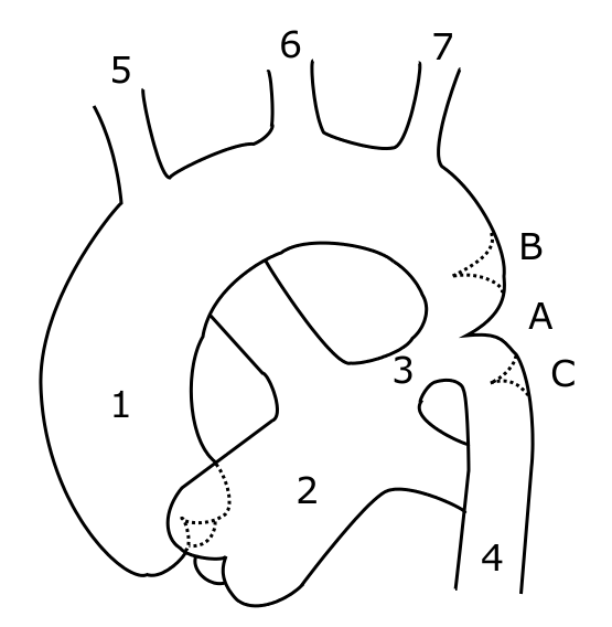
Schematic drawing of alternative locations of a coarctation of the aorta, relative to the ductus arteriosus. A: ductal coarctation, B: preductal coarctation, C: postductal coarctation. 1: Aorta ascendens, 2: Arteria pulmonalis, 3: Ductus arteriosus, 4: Aorta descendens, 5: Trunchus brachiocephalicus, 6: Arteria carotis communis sinister, 7: Arteria subclavia sinister
--------------------------------------------------------------------------
| ICD-10 | M31.4 |
|---|---|
| ICD-9 | 446.7 |
| OMIM | 207600 |
| DiseasesDB | 12879 |
| MedlinePlus | 001250 |
| eMedicine | radio/51 |
Takayasu's arteritis is an inflammatory disease of unknown etiology that affects the aorta and its branches. Although it has been reported worldwide, it shows a predilection for young Asian women. Females with this disease outnumber males by 8:1, and the age of onset is typically between 15 and 30 yr. In the Western world, atherosclerosis is a more frequent cause of obstruction of the aortic arch vessels than is Takayasu's arteritis.
Symptoms
About half of all patients develop an initial systemic illness with symptoms of malaise, fever, night sweats, weight loss, arthralgia, and fatigue. There is often an anemia and marked elevation of the ESR. This phase gradually subsides and is followed by a more chronic stage characterized by inflammatory and obliterative changes in the aorta and its branches. The other half of patients with Takayasu's arteritis present with only late vascular changes, without an antecedent systemic illness. In the late stage, weakness of the arterial walls may give rise to localized aneurysms. the disease can present in 4 forms namely, type1:localised to aortic arch and branches. type2:localised to descending thoracic aorta and abdominal aorta. type3:combination of types 1 and 2. type4:localised to pulmonary artery.
[edit] Treatments
The great majority of patients with Takayasu’s arteritis respond to prednisone. The usual starting dose is approximately 1 milligram per kilogram of body weight per day (for most people, this is approximately 60 milligrams a day). Because of the significant side effects of long-term high–dose prednisone use, the starting dose is tapered over several weeks to a dose that the physician feels is tolerable for the patient.
--------------------------------------------------------------------------
Kawasaki disease, also known as mucocutaneous lymph node syndrome, mucocutaneous lymph node disease, infantile polyarteritis and Kawasaki syndrome, is a poorly understood self-limited vasculitis that affects many organs, including the skin and mucous membranes, lymph nodes, blood vessel walls, and the heart. It does not seem to be contagious. It was first described in 1967 by Dr. Tomisaku Kawasaki in Japan.
ncidence, causes, and risk factors
By far, the highest incidence of Kawasaki disease occurs in Japan, though its incidence in the United States is increasing. Kawasaki disease is predominantly a disease of young children, with 80% of patients younger than 5 years of age. Additional risk factors in the United States include Asian race and male sex.
The causative agent of Kawasaki disease is still unknown. However, current etiological theories center primarily on immunological causes for the disease. Much research is being performed to discover a definitive toxin or antigenic substance, possibly a superantigen, that is the specific cause of the disease. An unknown virus may play a role as an inciting factor as well.
The cardiac complications are, by far, the most important aspect of the disease. Kawasaki disease can cause vasculitic changes (inflammation of blood vessels) in the coronary arteries and subsequent coronary artery aneurysms. These aneurysms can lead to myocardial infarction (heart attack) even in young children. Overall, about 10–18% of children with Kawasaki disease develop coronary artery anuerysms[1], with much higher prevalence among patients who are not treated early in the course of illness. Kawasaki disease is the most common cause of acquired heart disease among children in the United States.
Kawasaki disease often begins with a high and persistent fever that is not very responsive to normal doses of acetaminophen or ibuprofen. The fever may persist steadily for up to two weeks and is normally accompanied by irritability. Affected children develop red eyes, red mucous membranes in the mouth, red cracked lips, a "strawberry tongue" and swollen lymph nodes. Skin rashes occur early in the disease and peeling of the skin in the genital area, hands, and feet (especially around the nails and on the palms and soles) may occur in later phases. Some of these symptoms may come and go during the course of the illness. If left untreated, the symptoms will eventually relent but coronary artery aneurysms will not improve, resulting in a significant risk of death or disability due to myocardial infarction (heart attack). If treated in a timely fashion, this risk can be mostly avoided and the course of illness cut short.
[edit] Symptoms
- High-grade fever (greater than 39 °C or 102 °F; often as high as 40 °C or 104 °F) that normally lasts for more than a week if left untreated.
- Red eyes (conjunctivitis) without pus or drainage, also known as "conjunctival injection"
- Bright red, chapped, or cracked lips
- Red mucous membranes in the mouth
- Strawberry tongue, white coating on the tongue or prominent red bumps (papillae) on the back of the tongue
- Red palms of the hands and the soles of the feet
- Swollen hands and feet
- Rash which may take many forms, but not vesicular (blister-like), on the trunk
- Swollen lymph nodes (frequently only one lymph node is swollen), particularly in the neck area
- Joint pain (arthralgia) and swelling, frequently symmetrical
- Irritability
- Tachycardia (rapid heart beat)
- Peeling palms and soles (later in the illness); peeling may begin around the nails
